 Add to Chrome
 Add to Firefox
 Add to Edge
Artificial Intelligence-Assisted Prostate Cancer Diagnosis for Reduced Use of Immunohistochemistry
Mar 31, 2025Prostate cancer diagnosis heavily relies on histopathological evaluation, which is subject to variability. While immunohistochemical staining (IHC) assists in distinguishing benign from malignant tissue, it involves increased work, higher costs, and diagnostic delays. Artificial intelligence (AI) presents a promising solution to reduce reliance on IHC by accurately classifying atypical glands and borderline morphologies in hematoxylin & eosin (H&E) stained tissue sections. In this study, we evaluated an AI model's ability to minimize IHC use without compromising diagnostic accuracy by retrospectively analyzing prostate core needle biopsies from routine diagnostics at three different pathology sites. These cohorts were composed exclusively of difficult cases where the diagnosing pathologists required IHC to finalize the diagnosis. The AI model demonstrated area under the curve values of 0.951-0.993 for detecting cancer in routine H&E-stained slides. Applying sensitivity-prioritized diagnostic thresholds reduced the need for IHC staining by 44.4%, 42.0%, and 20.7% in the three cohorts investigated, without a single false negative prediction. This AI model shows potential for optimizing IHC use, streamlining decision-making in prostate pathology, and alleviating resource burdens.
The impact of tissue detection on diagnostic artificial intelligence algorithms in digital pathology
Mar 29, 2025
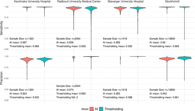


Tissue detection is a crucial first step in most digital pathology applications. Details of the segmentation algorithm are rarely reported, and there is a lack of studies investigating the downstream effects of a poor segmentation algorithm. Disregarding tissue detection quality could create a bottleneck for downstream performance and jeopardize patient safety if diagnostically relevant parts of the specimen are excluded from analysis in clinical applications. This study aims to determine whether performance of downstream tasks is sensitive to the tissue detection method, and to compare performance of classical and AI-based tissue detection. To this end, we trained an AI model for Gleason grading of prostate cancer in whole slide images (WSIs) using two different tissue detection algorithms: thresholding (classical) and UNet++ (AI). A total of 33,823 WSIs scanned on five digital pathology scanners were used to train the tissue detection AI model. The downstream Gleason grading algorithm was trained and tested using 70,524 WSIs from 13 clinical sites scanned on 13 different scanners. There was a decrease from 116 (0.43%) to 22 (0.08%) fully undetected tissue samples when switching from thresholding-based tissue detection to AI-based, suggesting an AI model may be more reliable than a classical model for avoiding total failures on slides with unusual appearance. On the slides where tissue could be detected by both algorithms, no significant difference in overall Gleason grading performance was observed. However, tissue detection dependent clinically significant variations in AI grading were observed in 3.5% of malignant slides, highlighting the importance of robust tissue detection for optimal clinical performance of diagnostic AI.
Self-Contrastive Weakly Supervised Learning Framework for Prognostic Prediction Using Whole Slide Images
May 24, 2024


We present a pioneering investigation into the application of deep learning techniques to analyze histopathological images for addressing the substantial challenge of automated prognostic prediction. Prognostic prediction poses a unique challenge as the ground truth labels are inherently weak, and the model must anticipate future events that are not directly observable in the image. To address this challenge, we propose a novel three-part framework comprising of a convolutional network based tissue segmentation algorithm for region of interest delineation, a contrastive learning module for feature extraction, and a nested multiple instance learning classification module. Our study explores the significance of various regions of interest within the histopathological slides and exploits diverse learning scenarios. The pipeline is initially validated on artificially generated data and a simpler diagnostic task. Transitioning to prognostic prediction, tasks become more challenging. Employing bladder cancer as use case, our best models yield an AUC of 0.721 and 0.678 for recurrence and treatment outcome prediction respectively.
NMGrad: Advancing Histopathological Bladder Cancer Grading with Weakly Supervised Deep Learning
May 24, 2024

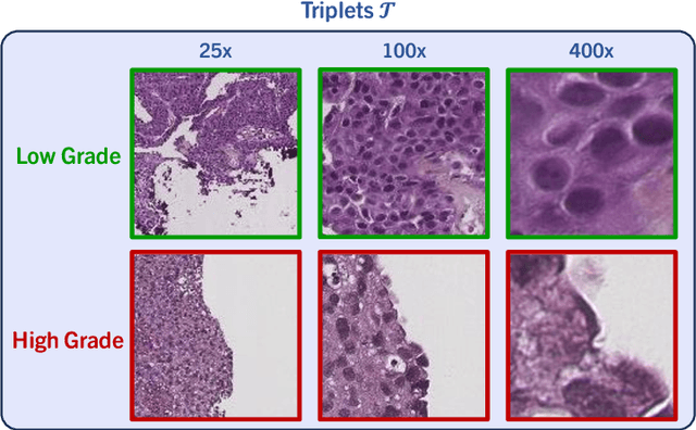

The most prevalent form of bladder cancer is urothelial carcinoma, characterized by a high recurrence rate and substantial lifetime treatment costs for patients. Grading is a prime factor for patient risk stratification, although it suffers from inconsistencies and variations among pathologists. Moreover, absence of annotations in medical imaging difficults training deep learning models. To address these challenges, we introduce a pipeline designed for bladder cancer grading using histological slides. First, it extracts urothelium tissue tiles at different magnification levels, employing a convolutional neural network for processing for feature extraction. Then, it engages in the slide-level prediction process. It employs a nested multiple instance learning approach with attention to predict the grade. To distinguish different levels of malignancy within specific regions of the slide, we include the origins of the tiles in our analysis. The attention scores at region level is shown to correlate with verified high-grade regions, giving some explainability to the model. Clinical evaluations demonstrate that our model consistently outperforms previous state-of-the-art methods.
Equipping Computational Pathology Systems with Artifact Processing Pipelines: A Showcase for Computation and Performance Trade-offs
Mar 13, 2024Histopathology is a gold standard for cancer diagnosis under a microscopic examination. However, histological tissue processing procedures result in artifacts, which are ultimately transferred to the digitized version of glass slides, known as whole slide images (WSIs). Artifacts are diagnostically irrelevant areas and may result in wrong deep learning (DL) algorithms predictions. Therefore, detecting and excluding artifacts in the computational pathology (CPATH) system is essential for reliable automated diagnosis. In this paper, we propose a mixture of experts (MoE) scheme for detecting five notable artifacts, including damaged tissue, blur, folded tissue, air bubbles, and histologically irrelevant blood from WSIs. First, we train independent binary DL models as experts to capture particular artifact morphology. Then, we ensemble their predictions using a fusion mechanism. We apply probabilistic thresholding over the final probability distribution to improve the sensitivity of the MoE. We developed DL pipelines using two MoEs and two multiclass models of state-of-the-art deep convolutional neural networks (DCNNs) and vision transformers (ViTs). DCNNs-based MoE and ViTs-based MoE schemes outperformed simpler multiclass models and were tested on datasets from different hospitals and cancer types, where MoE using DCNNs yielded the best results. The proposed MoE yields 86.15% F1 and 97.93% sensitivity scores on unseen data, retaining less computational cost for inference than MoE using ViTs. This best performance of MoEs comes with relatively higher computational trade-offs than multiclass models. The proposed artifact detection pipeline will not only ensure reliable CPATH predictions but may also provide quality control.
A Dual Convolutional Neural Network Pipeline for Melanoma Diagnostics and Prognostics
Dec 14, 2023Melanoma is a type of cancer that begins in the cells controlling the pigment of the skin, and it is often referred to as the most dangerous skin cancer. Diagnosing melanoma can be time-consuming, and a recent increase in melanoma incidents indicates a growing demand for a more efficient diagnostic process. This paper presents a pipeline for melanoma diagnostics, leveraging two convolutional neural networks, a diagnosis, and a prognosis model. The diagnostic model is responsible for localizing malignant patches across whole slide images and delivering a patient-level diagnosis as malignant or benign. Further, the prognosis model utilizes the diagnostic model's output to provide a patient-level prognosis as good or bad. The full pipeline has an F1 score of 0.79 when tested on data from the same distribution as it was trained on.
Balancing Privacy and Progress in Artificial Intelligence: Anonymization in Histopathology for Biomedical Research and Education
Aug 08, 2023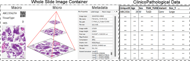
The advancement of biomedical research heavily relies on access to large amounts of medical data. In the case of histopathology, Whole Slide Images (WSI) and clinicopathological information are valuable for developing Artificial Intelligence (AI) algorithms for Digital Pathology (DP). Transferring medical data "as open as possible" enhances the usability of the data for secondary purposes but poses a risk to patient privacy. At the same time, existing regulations push towards keeping medical data "as closed as necessary" to avoid re-identification risks. Generally, these legal regulations require the removal of sensitive data but do not consider the possibility of data linkage attacks due to modern image-matching algorithms. In addition, the lack of standardization in DP makes it harder to establish a single solution for all formats of WSIs. These challenges raise problems for bio-informatics researchers in balancing privacy and progress while developing AI algorithms. This paper explores the legal regulations and terminologies for medical data-sharing. We review existing approaches and highlight challenges from the histopathological perspective. We also present a data-sharing guideline for histological data to foster multidisciplinary research and education.
Deep Learning for Predicting Metastasis on Melanoma WSIs
Mar 10, 2023


Northern Europe has the second highest mortality rate of melanoma globally. In 2020, the mortality rate of melanoma rose to 1.9 per 100 000 habitants. Melanoma prognosis is based on a pathologist's subjective visual analysis of the patient's tumor. This methodology is heavily time-consuming, and the prognosis variability among experts is notable, drastically jeopardizing its reproducibility. Thus, the need for faster and more reproducible methods arises. Machine learning has paved its way into digital pathology, but so far, most contributions are on localization, segmentation, and diagnostics, with little emphasis on prognostics. This paper presents a convolutional neural network (CNN) method based on VGG16 to predict melanoma prognosis as the presence of metastasis within five years. Patches are extracted from regions of interest from Whole Slide Images (WSIs) at different magnification levels used in model training and validation. Results infer that utilizing WSI patches at 20x magnification level has the best performance, with an F1 score of 0.7667 and an AUC of 0.81.
Detection and Localization of Melanoma Skin Cancer in Histopathological Whole Slide Images
Feb 24, 2023


Melanoma diagnosed and treated in its early stages can increase the survival rate. A projected increase in skin cancer incidents and a dearth of dermatopathologists have emphasized the need for computational pathology (CPATH) systems. CPATH systems with deep learning (DL) models have the potential to identify the presence of melanoma by exploiting underlying morphological and cellular features. This paper proposes a DL method to detect melanoma and distinguish between normal skin and benign/malignant melanocytic lesions in Whole Slide Images (WSI). Our method detects lesions with high accuracy and localizes them on a WSI to identify potential regions of interest for pathologists. Interestingly, our DL method relies on using a single CNN network to create localization maps first and use them to perform slide-level predictions to determine patients who have melanoma. Our best model provides favorable patch-wise classification results with a 0.992 F1 score and 0.99 sensitivity on unseen data. The source code is https://github.com/RogerAmundsen/Melanoma-Diagnosis-and-Localization-from-Whole-Slide-Images-using-Convolutional-Neural-Networks.