Deepika Sirohi
CLASS-M: Adaptive stain separation-based contrastive learning with pseudo-labeling for histopathological image classification
Jan 04, 2024


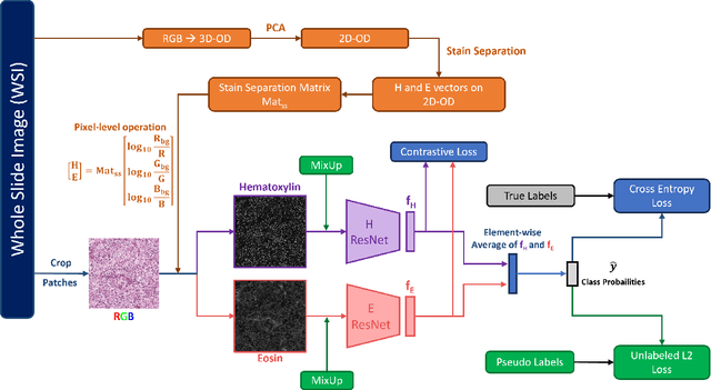
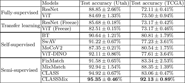
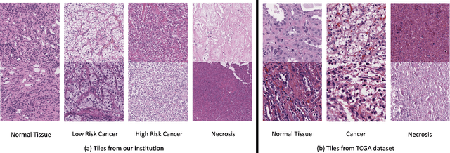
Abstract:Histopathological image classification is an important task in medical image analysis. Recent approaches generally rely on weakly supervised learning due to the ease of acquiring case-level labels from pathology reports. However, patch-level classification is preferable in applications where only a limited number of cases are available or when local prediction accuracy is critical. On the other hand, acquiring extensive datasets with localized labels for training is not feasible. In this paper, we propose a semi-supervised patch-level histopathological image classification model, named CLASS-M, that does not require extensively labeled datasets. CLASS-M is formed by two main parts: a contrastive learning module that uses separated Hematoxylin and Eosin images generated through an adaptive stain separation process, and a module with pseudo-labels using MixUp. We compare our model with other state-of-the-art models on two clear cell renal cell carcinoma datasets. We demonstrate that our CLASS-M model has the best performance on both datasets. Our code is available at github.com/BzhangURU/Paper_CLASS-M/tree/main
A Pathologist-Informed Workflow for Classification of Prostate Glands in Histopathology
Sep 27, 2022Abstract:Pathologists diagnose and grade prostate cancer by examining tissue from needle biopsies on glass slides. The cancer's severity and risk of metastasis are determined by the Gleason grade, a score based on the organization and morphology of prostate cancer glands. For diagnostic work-up, pathologists first locate glands in the whole biopsy core, and -- if they detect cancer -- they assign a Gleason grade. This time-consuming process is subject to errors and significant inter-observer variability, despite strict diagnostic criteria. This paper proposes an automated workflow that follows pathologists' \textit{modus operandi}, isolating and classifying multi-scale patches of individual glands in whole slide images (WSI) of biopsy tissues using distinct steps: (1) two fully convolutional networks segment epithelium versus stroma and gland boundaries, respectively; (2) a classifier network separates benign from cancer glands at high magnification; and (3) an additional classifier predicts the grade of each cancer gland at low magnification. Altogether, this process provides a gland-specific approach for prostate cancer grading that we compare against other machine-learning-based grading methods.
* Published as a workshop paper at MICCAI MOVI 2022
Stain based contrastive co-training for histopathological image analysis
Jun 24, 2022


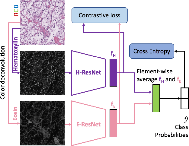
Abstract:We propose a novel semi-supervised learning approach for classification of histopathology images. We employ strong supervision with patch-level annotations combined with a novel co-training loss to create a semi-supervised learning framework. Co-training relies on multiple conditionally independent and sufficient views of the data. We separate the hematoxylin and eosin channels in pathology images using color deconvolution to create two views of each slide that can partially fulfill these requirements. Two separate CNNs are used to embed the two views into a joint feature space. We use a contrastive loss between the views in this feature space to implement co-training. We evaluate our approach in clear cell renal cell and prostate carcinomas, and demonstrate improvement over state-of-the-art semi-supervised learning methods.
 Add to Chrome
 Add to Firefox
 Add to Edge