Christoph Leitner
PuLsE: Accurate and Robust Ultrasound-based Continuous Heart-Rate Monitoring on a Wrist-Worn IoT Device
Oct 21, 2024


Abstract:This work explores the feasibility of employing ultrasound (US) US technology in a wrist-worn IoT device for low-power, high-fidelity heart-rate (HR) extraction. US offers deep tissue penetration and can monitor pulsatile arterial blood flow in large vessels and the surrounding tissue, potentially improving robustness and accuracy compared to PPG. We present an IoT wearable system prototype utilizing a commercial microcontroller MCU employing the onboard ADC to capture high frequency US signals and an innovative low-power US pulser. An envelope filter lowers the bandwidth of the US signal by a factor of >5x, reducing the system's acquisition requirements without compromising accuracy (correlation coefficient between HR extracted from enveloped and raw signals, r(92)=0.99, p<0.001). The full signal processing pipeline is ported to fixed point arithmetic for increased energy efficiency and runs entirely onboard. The system has an average power consumption of 5.8mW, competitive with PPG based systems, and the HR extraction algorithm requires only 68kB of RAM and 71ms of processing time on an ARM Cortex-M4 MCU. The system is estimated to run continuously for more than 7 days on a smartwatch battery. To accurately evaluate the proposed circuit and algorithm and identify the anatomical location on the wrist with the highest accuracy for HR extraction, we collected a dataset from 10 healthy adults at three different wrist positions. The dataset comprises roughly 5 hours of HR data with an average of 80.6+-16.3 bpm. During recording, we synchronized the established ECG gold standard with our US-based method. The comparisons yields a Pearson correlation coefficient of r(92)=0.99, p<0.001 and a mean error of 0.69+-1.99 bpm in the lateral wrist position near the radial artery. The dataset and code have been open-sourced at https://github.com/mgiordy/Ultrasound-Heart-Rate
Towards a Novel Ultrasound System Based on Low-Frequency Feature Extraction From a Fully-Printed Flexible Transducer
Sep 25, 2023Abstract:Ultrasound is a key technology in healthcare, and it is being explored for non-invasive, wearable, continuous monitoring of vital signs. However, its widespread adoption in this scenario is still hindered by the size, complexity, and power consumption of current devices. Moreover, such an application demands adaptability to human anatomy, which is hard to achieve with current transducer technology. This paper presents a novel ultrasound system prototype based on a fully printed, lead-free, and flexible polymer ultrasound transducer, whose bending radius promises good adaptability to the human anatomy. Our application scenario focuses on continuous blood flow monitoring. We implemented a hardware envelope filter to efficiently transpose high-frequency ultrasound signals to a lower-frequency spectrum. This reduces computational and power demands with little to no degradation in the task proposed for this work. We validated our method on a setup that mimics human blood flow by using a flow phantom and a peristaltic pump simulating 3 different heartbeat rhythms: 60, 90 and 120 beats per minute. Our ultrasound setup reconstructs peristaltic pump frequencies with errors of less than 0.05 Hz (3 bpm) from the set pump frequency, both for the raw echo and the enveloped echo. The analog pre-processing showed a promising reduction of signal bandwidth of more than 6x: pulse-echo signals of transducers excited at 12.5 MHz were reduced to about 2 MHz. Thus, allowing consumer MCUs to acquire and elaborate signals within mW-power range in an inexpensive fashion.
Skilog: A Smart Sensor System for Performance Analysis and Biofeedback in Ski Jumping
Sep 25, 2023


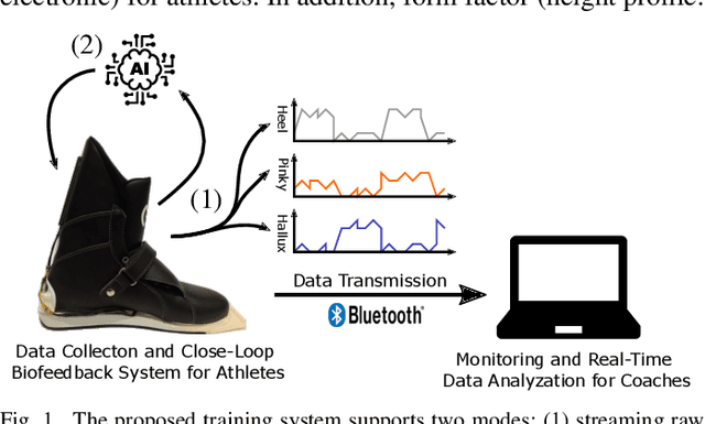
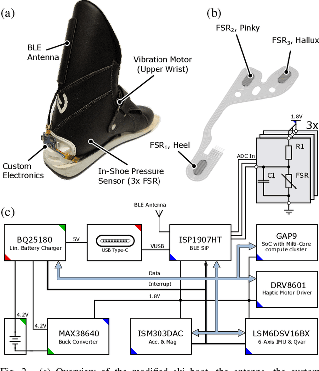
Abstract:In ski jumping, low repetition rates of jumps limit the effectiveness of training. Thus, increasing learning rate within every single jump is key to success. A critical element of athlete training is motor learning, which has been shown to be accelerated by feedback methods. In particular, a fine-grained control of the center of gravity in the in-run is essential. This is because the actual takeoff occurs within a blink of an eye ($\sim$300ms), thus any unbalanced body posture during the in-run will affect flight. This paper presents a smart, compact, and energy-efficient wireless sensor system for real-time performance analysis and biofeedback during ski jumping. The system operates by gauging foot pressures at three distinct points on the insoles of the ski boot at 100Hz. Foot pressure data can either be directly sent to coaches to improve their feedback, or fed into a ML model to give athletes instantaneous in-action feedback using a vibration motor in the ski boot. In the biofeedback scenario, foot pressures act as input variables for an optimized XGBoost model. We achieve a high predictive accuracy of 92.7% for center of mass predictions (dorsal shift, neutral stand, ventral shift). Subsequently, we parallelized and fine-tuned our XGBoost model for a RISC-V based low power parallel processor (GAP9), based on the PULP architecture. We demonstrate real-time detection and feedback (0.0109ms/inference) using our on-chip deployment. The proposed smart system is unobtrusive with a slim form factor (13mm baseboard, 3.2mm antenna) and a lightweight build (26g). Power consumption analysis reveals that the system's energy-efficient design enables sustained operation over multiple days (up to 300 hours) without requiring recharge.
A Wearable Ultra-Low-Power sEMG-Triggered Ultrasound System for Long-Term Muscle Activity Monitoring
Sep 14, 2023Abstract:Surface electromyography (sEMG) is a well-established approach to monitor muscular activity on wearable and resource-constrained devices. However, when measuring deeper muscles, its low signal-to-noise ratio (SNR), high signal attenuation, and crosstalk degrade sensing performance. Ultrasound (US) complements sEMG effectively with its higher SNR at high penetration depths. In fact, combining US and sEMG improves the accuracy of muscle dynamic assessment, compared to using only one modality. However, the power envelope of US hardware is considerably higher than that of sEMG, thus inflating energy consumption and reducing the battery life. This work proposes a wearable solution that integrates both modalities and utilizes an EMG-driven wake-up approach to achieve ultra-low power consumption as needed for wearable long-term monitoring. We integrate two wearable state-of-the-art (SoA) US and ExG biosignal acquisition devices to acquire time-synchronized measurements of the short head of the biceps. To minimize power consumption, the US probe is kept in a sleep state when there is no muscle activity. sEMG data are processed on the probe (filtering, envelope extraction and thresholding) to identify muscle activity and generate a trigger to wake-up the US counterpart. The US acquisition starts before muscle fascicles displacement thanks to a triggering time faster than the electromechanical delay (30-100 ms) between the neuromuscular junction stimulation and the muscle contraction. Assuming a muscle contraction of 200 ms at a contraction rate of 1 Hz, the proposed approach enables more than 59% energy saving (with a full-system average power consumption of 12.2 mW) as compared to operating both sEMG and US continuously.
A Human-Centered Machine-Learning Approach for Muscle-Tendon Junction Tracking in Ultrasound Images
Feb 10, 2022


Abstract:Biomechanical and clinical gait research observes muscles and tendons in limbs to study their functions and behaviour. Therefore, movements of distinct anatomical landmarks, such as muscle-tendon junctions, are frequently measured. We propose a reliable and time efficient machine-learning approach to track these junctions in ultrasound videos and support clinical biomechanists in gait analysis. In order to facilitate this process, a method based on deep-learning was introduced. We gathered an extensive dataset, covering 3 functional movements, 2 muscles, collected on 123 healthy and 38 impaired subjects with 3 different ultrasound systems, and providing a total of 66864 annotated ultrasound images in our network training. Furthermore, we used data collected across independent laboratories and curated by researchers with varying levels of experience. For the evaluation of our method a diverse test-set was selected that is independently verified by four specialists. We show that our model achieves similar performance scores to the four human specialists in identifying the muscle-tendon junction position. Our method provides time-efficient tracking of muscle-tendon junctions, with prediction times of up to 0.078 seconds per frame (approx. 100 times faster than manual labeling). All our codes, trained models and test-set were made publicly available and our model is provided as a free-to-use online service on https://deepmtj.org/.
Automatic Tracking of the Muscle Tendon Junction in Healthy and Impaired Subjects using Deep Learning
May 05, 2020


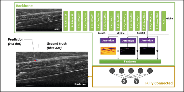
Abstract:Recording muscle tendon junction displacements during movement, allows separate investigation of the muscle and tendon behaviour, respectively. In order to provide a fully-automatic tracking method, we employ a novel deep learning approach to detect the position of the muscle tendon junction in ultrasound images. We utilize the attention mechanism to enable the network to focus on relevant regions and to obtain a better interpretation of the results. Our data set consists of a large cohort of 79 healthy subjects and 28 subjects with movement limitations performing passive full range of motion and maximum contraction movements. Our trained network shows robust detection of the muscle tendon junction on a diverse data set of varying quality with a mean absolute error of 2.55$\pm$1 mm. We show that our approach can be applied for various subjects and can be operated in real-time. The complete software package is available for open-source use via: https://github.com/luuleitner/deepMTJ
 Add to Chrome
 Add to Firefox
 Add to Edge