Christine U. Lee
Radiology Department at Mayo Clinic, Rochester MN
A Radiological Clip Design Using Ultrasound Identification to Improve Localization
Aug 30, 2023


Abstract:Objective: We demonstrate the use of ultrasound to receive an acoustic signal transmitted from a radiological clip designed from a custom circuit. This signal encodes an identification number and is localized and identified wirelessly by the ultrasound imaging system. Methods: We designed and constructed the test platform with a Teensy 4.0 microcontroller core to detect ultrasonic imaging pulses received by a transducer embedded in a phantom, which acted as the radiological clip. Ultrasound identification (USID) signals were generated and transmitted as a result. The phantom and clip were imaged using an ultrasonic array (Philips L7-4) connected to a Verasonics Vantage 128 system operating in pulse inversion (PI) mode. Cross-correlations were performed to localize and identify the code sequences in the PI images. Results: USID signals were detected and visualized on B-mode images of the phantoms with up to sub-millimeter localization accuracy. The average detection rate across 1,600 frames of ultrasound data was 94.6%. Tested ID values exhibited differences in detection rates. Conclusion: The USID clip produced identifiable, distinguishable, and localizable signals when imaged. Significance: Radiological clips are used to mark breast cancer being treated by neoadjuvant chemotherapy (NAC) via implant in or near treated lesions. As NAC progresses, available marking clips can lose visibility in ultrasound, the imaging modality of choice for monitoring NAC-treated lesions. By transmitting an active signal, more accurate and reliable ultrasound localization of these clips could be achieved and multiple clips with different ID values could be imaged in the same field of view.
Joint localization and classification of breast tumors on ultrasound images using a novel auxiliary attention-based framework
Oct 11, 2022


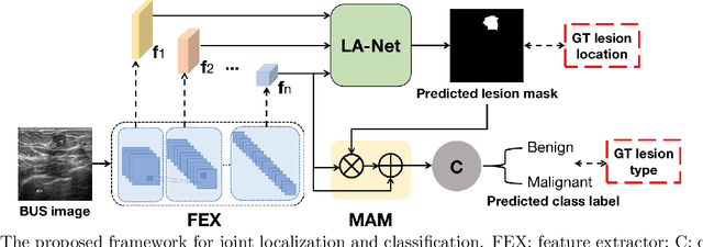
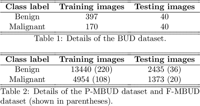
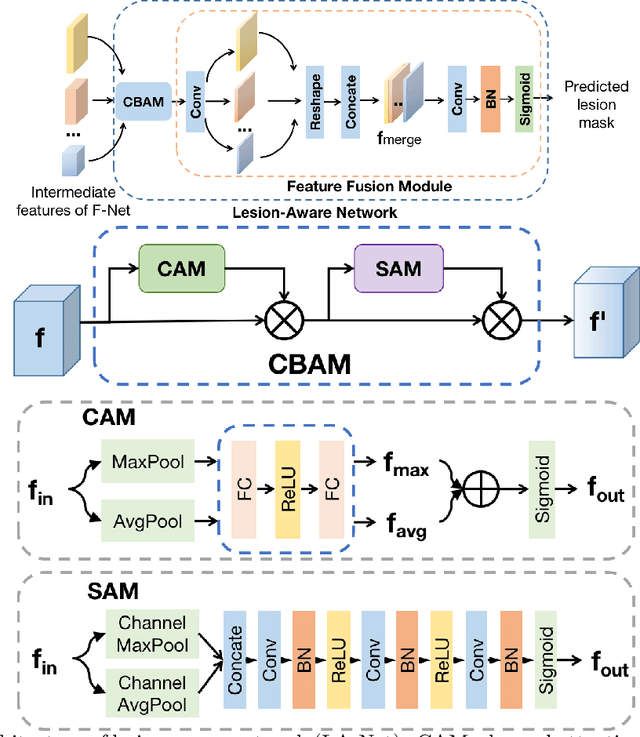
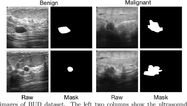
Abstract:Automatic breast lesion detection and classification is an important task in computer-aided diagnosis, in which breast ultrasound (BUS) imaging is a common and frequently used screening tool. Recently, a number of deep learning-based methods have been proposed for joint localization and classification of breast lesions using BUS images. In these methods, features extracted by a shared network trunk are appended by two independent network branches to achieve classification and localization. Improper information sharing might cause conflicts in feature optimization in the two branches and leads to performance degradation. Also, these methods generally require large amounts of pixel-level annotated data for model training. To overcome these limitations, we proposed a novel joint localization and classification model based on the attention mechanism and disentangled semi-supervised learning strategy. The model used in this study is composed of a classification network and an auxiliary lesion-aware network. By use of the attention mechanism, the auxiliary lesion-aware network can optimize multi-scale intermediate feature maps and extract rich semantic information to improve classification and localization performance. The disentangled semi-supervised learning strategy only requires incomplete training datasets for model training. The proposed modularized framework allows flexible network replacement to be generalized for various applications. Experimental results on two different breast ultrasound image datasets demonstrate the effectiveness of the proposed method. The impacts of various network factors on model performance are also investigated to gain deep insights into the designed framework.
 Add to Chrome
 Add to Firefox
 Add to Edge