Alvin Chen
Ultrasound Image Synthesis Using Generative AI for Lung Ultrasound Detection
Jan 10, 2025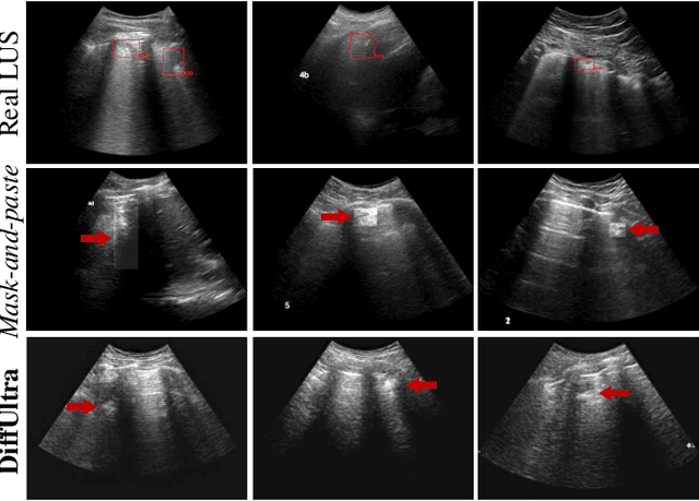

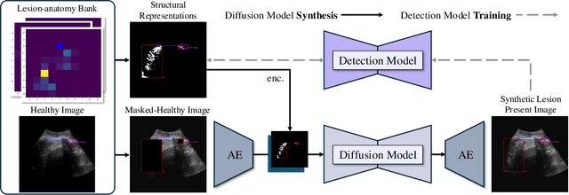

Abstract:Developing reliable healthcare AI models requires training with representative and diverse data. In imbalanced datasets, model performance tends to plateau on the more prevalent classes while remaining low on less common cases. To overcome this limitation, we propose DiffUltra, the first generative AI technique capable of synthesizing realistic Lung Ultrasound (LUS) images with extensive lesion variability. Specifically, we condition the generative AI by the introduced Lesion-anatomy Bank, which captures the lesion's structural and positional properties from real patient data to guide the image synthesis.We demonstrate that DiffUltra improves consolidation detection by 5.6% in AP compared to the models trained solely on real patient data. More importantly, DiffUltra increases data diversity and prevalence of rare cases, leading to a 25% AP improvement in detecting rare instances such as large lung consolidations, which make up only 10% of the dataset.
Contrastive Self-Supervised Learning for Spatio-Temporal Analysis of Lung Ultrasound Videos
Oct 14, 2023Abstract:Self-supervised learning (SSL) methods have shown promise for medical imaging applications by learning meaningful visual representations, even when the amount of labeled data is limited. Here, we extend state-of-the-art contrastive learning SSL methods to 2D+time medical ultrasound video data by introducing a modified encoder and augmentation method capable of learning meaningful spatio-temporal representations, without requiring constraints on the input data. We evaluate our method on the challenging clinical task of identifying lung consolidations (an important pathological feature) in ultrasound videos. Using a multi-center dataset of over 27k lung ultrasound videos acquired from over 500 patients, we show that our method can significantly improve performance on downstream localization and classification of lung consolidation. Comparisons against baseline models trained without SSL show that the proposed methods are particularly advantageous when the size of labeled training data is limited (e.g., as little as 5% of the training set).
Weakly Semi-Supervised Detection in Lung Ultrasound Videos
Aug 08, 2023Abstract:Frame-by-frame annotation of bounding boxes by clinical experts is often required to train fully supervised object detection models on medical video data. We propose a method for improving object detection in medical videos through weak supervision from video-level labels. More concretely, we aggregate individual detection predictions into video-level predictions and extend a teacher-student training strategy to provide additional supervision via a video-level loss. We also introduce improvements to the underlying teacher-student framework, including methods to improve the quality of pseudo-labels based on weak supervision and adaptive schemes to optimize knowledge transfer between the student and teacher networks. We apply this approach to the clinically important task of detecting lung consolidations (seen in respiratory infections such as COVID-19 pneumonia) in medical ultrasound videos. Experiments reveal that our framework improves detection accuracy and robustness compared to baseline semi-supervised models, and improves efficiency in data and annotation usage.
Automatic ultrasound vessel segmentation with deep spatiotemporal context learning
Nov 03, 2021
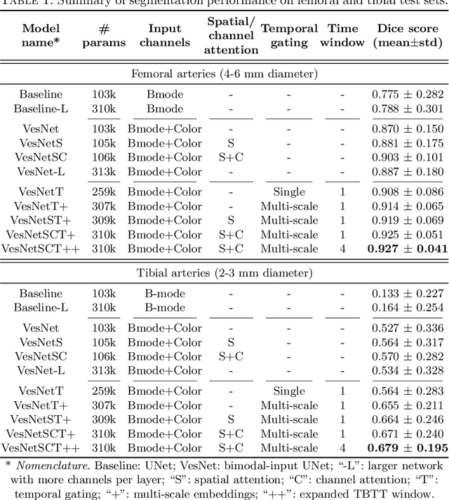
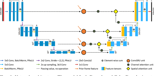
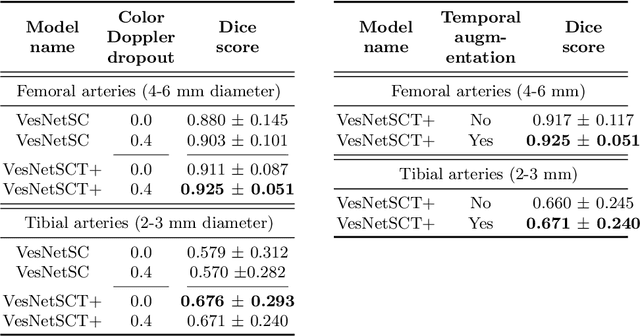
Abstract:Accurate, real-time segmentation of vessel structures in ultrasound image sequences can aid in the measurement of lumen diameters and assessment of vascular diseases. This, however, remains a challenging task, particularly for extremely small vessels that are difficult to visualize. We propose to leverage the rich spatiotemporal context available in ultrasound to improve segmentation of small-scale lower-extremity arterial vasculature. We describe efficient deep learning methods that incorporate temporal, spatial, and feature-aware contextual embeddings at multiple resolution scales while jointly utilizing information from B-mode and Color Doppler signals. Evaluating on femoral and tibial artery scans performed on healthy subjects by an expert ultrasonographer, and comparing to consensus expert ground-truth annotations of inner lumen boundaries, we demonstrate real-time segmentation using the context-aware models and show that they significantly outperform comparable baseline approaches.
 Add to Chrome
 Add to Firefox
 Add to Edge