Alessandro Sbrizzi
Quantitative mapping from conventional MRI using self-supervised physics-guided deep learning: applications to a large-scale, clinically heterogeneous dataset
Jan 08, 2026Abstract:Magnetic resonance imaging (MRI) is a cornerstone of clinical neuroimaging, yet conventional MRIs provide qualitative information heavily dependent on scanner hardware and acquisition settings. While quantitative MRI (qMRI) offers intrinsic tissue parameters, the requirement for specialized acquisition protocols and reconstruction algorithms restricts its availability and impedes large-scale biomarker research. This study presents a self-supervised physics-guided deep learning framework to infer quantitative T1, T2, and proton-density (PD) maps directly from widely available clinical conventional T1-weighted, T2-weighted, and FLAIR MRIs. The framework was trained and evaluated on a large-scale, clinically heterogeneous dataset comprising 4,121 scan sessions acquired at our institution over six years on four different 3 T MRI scanner systems, capturing real-world clinical variability. The framework integrates Bloch-based signal models directly into the training objective. Across more than 600 test sessions, the generated maps exhibited white matter and gray matter values consistent with literature ranges. Additionally, the generated maps showed invariance to scanner hardware and acquisition protocol groups, with inter-group coefficients of variation $\leq$ 1.1%. Subject-specific analyses demonstrated excellent voxel-wise reproducibility across scanner systems and sequence parameters, with Pearson $r$ and concordance correlation coefficients exceeding 0.82 for T1 and T2. Mean relative voxel-wise differences were low across all quantitative parameters, especially for T2 ($<$ 6%). These results indicate that the proposed framework can robustly transform diverse clinical conventional MRI data into quantitative maps, potentially paving the way for large-scale quantitative biomarker research.
Time-efficient, high-resolution 3T whole-brain relaxometry using Cartesian 3D MR-STAT with CSF suppression
Mar 22, 2024


Abstract:Purpose: Current 3D Magnetic Resonance Spin TomogrAphy in Time-domain (MR-STAT) protocols use transient-state, gradient-spoiled gradient-echo sequences that are prone to cerebrospinal fluid (CSF) pulsation artifacts when applied to the brain. This study aims at developing a 3D MR-STAT protocol for whole-brain relaxometry that overcomes the challenges posed by CSF-induced ghosting artifacts. Method: We optimized the flip-angle train within the Cartesian 3D MR-STAT framework to achieve two objectives: (1) minimization of the noise level in the reconstructed quantitative maps, and (2) reduction of the CSF-to-white-matter signal ratio to suppress CSF signal and the associated pulsation artifacts. The optimized new sequence was tested on a gel/water-phantom to evaluate the accuracy of the quantitative maps, and on healthy volunteers to explore the effectiveness of the CSF artifact suppression and robustness of the new protocol. Results: A new optimized sequence with both high parameter encoding capability and low CSF intensity was proposed and initially validated in the gel/water-phantom experiment. From in-vivo experiments with five volunteers, the proposed CSF-suppressed sequence shows no CSF ghosting artifacts and overall greatly improved image quality for all quantitative maps compared to the baseline sequence. Statistical analysis indicated low inter-subject and inter-scan variability for quantitative parameters in gray matter and white matter (1.6%-2.4% for T1 and 2.0%-4.6% for T2), demonstrating the robustness of the new sequence. Conclusion: We presented a new 3D MR-STAT sequence with CSF suppression that effectively eliminates CSF pulsation artifacts. The new sequence ensures consistently high-quality, 1mm^3 whole-brain relaxometry within a rapid 5.5-minute scan time.
Time-Resolved Reconstruction of Motion, Force, and Stiffness using Spectro-Dynamic MRI
Oct 11, 2023
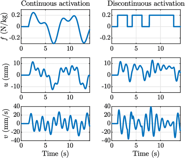
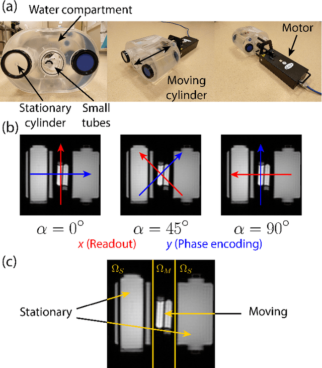

Abstract:Measuring the dynamics and mechanical properties of muscles and joints is important to understand the (patho)physiology of muscles. However, acquiring dynamic time-resolved MRI data is challenging. We have previously developed Spectro-Dynamic MRI which allows the characterization of dynamical systems at a high spatial and temporal resolution directly from k-space data. This work presents an extended Spectro-Dynamic MRI framework that reconstructs 1) time-resolved MR images, 2) time-resolved motion fields, 3) dynamical parameters, and 4) an activation force, at a temporal resolution of 11 ms. An iterative algorithm solves a minimization problem containing four terms: a motion model relating the motion to the fully-sampled k-space data, a dynamical model describing the expected type of dynamics, a data consistency term describing the undersampling pattern, and finally a regularization term for the activation force. We acquired MRI data using a dynamic motion phantom programmed to move like an actively driven linear elastic system, from which all dynamic variables could be accurately reconstructed, regardless of the sampling pattern. The proposed method performed better than a two-step approach, where time-resolved images were first reconstructed from the undersampled data without any information about the motion, followed by a motion estimation step.
Real-time myocardial landmark tracking for MRI-guided cardiac radio-ablation using Gaussian Processes
Jun 19, 2023Abstract:The high speed of cardiorespiratory motion introduces a unique challenge for cardiac stereotactic radio-ablation (STAR) treatments with the MR-linac. Such treatments require tracking myocardial landmarks with a maximum latency of 100 ms, which includes the acquisition of the required data. The aim of this study is to present a new method that allows to track myocardial landmarks from few readouts of MRI data, thereby achieving a latency sufficient for STAR treatments. We present a tracking framework that requires only few readouts of k-space data as input, which can be acquired at least an order of magnitude faster than MR-images. Combined with the real-time tracking speed of a probabilistic machine learning framework called Gaussian Processes, this allows to track myocardial landmarks with a sufficiently low latency for cardiac STAR guidance, including both the acquisition of required data, and the tracking inference. The framework is demonstrated in 2D on a motion phantom, and in vivo on volunteers and a ventricular tachycardia (arrhythmia) patient. Moreover, the feasibility of an extension to 3D was demonstrated by in silico 3D experiments with a digital motion phantom. The framework was compared with template matching - a reference, image-based, method - and linear regression methods. Results indicate an order of magnitude lower total latency (<10 ms) for the proposed framework in comparison with alternative methods. The root-mean-square-distances and mean end-point-distance with the reference tracking method was less than 0.8 mm for all experiments, showing excellent (sub-voxel) agreement. The high accuracy in combination with a total latency of less than 10 ms - including data acquisition and processing - make the proposed method a suitable candidate for tracking during STAR treatments.
Generalizable synthetic MRI with physics-informed convolutional networks
May 21, 2023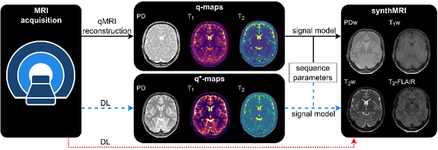

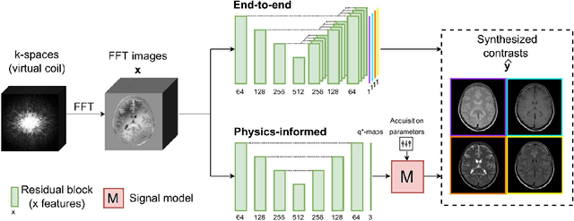

Abstract:In this study, we develop a physics-informed deep learning-based method to synthesize multiple brain magnetic resonance imaging (MRI) contrasts from a single five-minute acquisition and investigate its ability to generalize to arbitrary contrasts to accelerate neuroimaging protocols. A dataset of fifty-five subjects acquired with a standard MRI protocol and a five-minute transient-state sequence was used to develop a physics-informed deep learning-based method. The model, based on a generative adversarial network, maps data acquired from the five-minute scan to "effective" quantitative parameter maps, here named q*-maps, by using its generated PD, T1, and T2 values in a signal model to synthesize four standard contrasts (proton density-weighted, T1-weighted, T2-weighted, and T2-weighted fluid-attenuated inversion recovery), from which losses are computed. The q*-maps are compared to literature values and the synthetic contrasts are compared to an end-to-end deep learning-based method proposed by literature. The generalizability of the proposed method is investigated for five volunteers by synthesizing three non-standard contrasts unseen during training and comparing these to respective ground truth acquisitions via contrast-to-noise ratio and quantitative assessment. The physics-informed method was able to match the high-quality synthMRI of the end-to-end method for the four standard contrasts, with mean \pm standard deviation structural similarity metrics above 0.75 \pm 0.08 and peak signal-to-noise ratios above 22.4 \pm 1.9 and 22.6 \pm 2.1. Additionally, the physics-informed method provided retrospective contrast adjustment, with visually similar signal contrast and comparable contrast-to-noise ratios to the ground truth acquisitions for three sequences unused for model training, demonstrating its generalizability and potential application to accelerate neuroimaging protocols.
Towards retrospective motion correction and reconstruction for clinical 3D brain MRI protocols with a reference contrast
Jan 03, 2023Abstract:Motion artifacts often spoil the radiological interpretation of MR images, and in the most severe cases the scan needs be repeated, with additional costs for the provider. We discuss the application of a novel 3D retrospective rigid motion correction and reconstruction scheme for MRI, which leverages multiple scans contained in a MR session. Typically, in a multi-contrast MR session, motion does not equally affect all the scans, and some motion-free scans are generally available, so that we can exploit their anatomic similarity. The uncorrupted scan is used as a reference in a generalized rigid-motion registration problem to remove the motion artifacts affecting the corrupted scans. We discuss the potential of the proposed algorithm with a prospective in-vivo study and clinical 3D brain protocols. This framework can be easily incorporated into the existing clinical practice with no disruption to the conventional workflow.
Acceleration Strategies for MR-STAT: Achieving High-Resolution Reconstructions on a Desktop PC within 3 minutes
May 04, 2022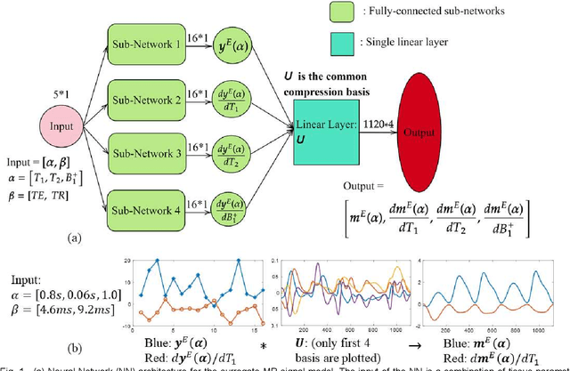
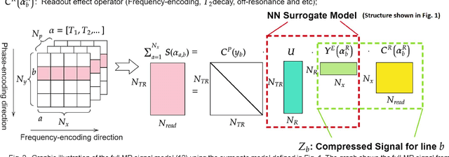
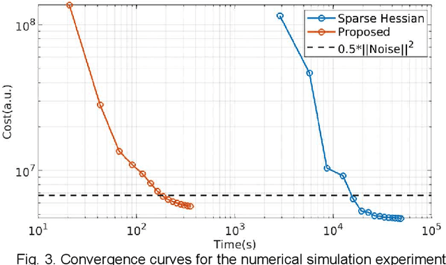
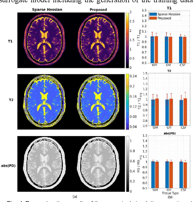
Abstract:MR-STAT is an emerging quantitative magnetic resonance imaging technique which aims at obtaining multi-parametric tissue parameter maps from single short scans. It describes the relationship between the spatial-domain tissue parameters and the time-domain measured signal by using a comprehensive, volumetric forward model. The MR-STAT reconstruction solves a large-scale nonlinear problem, thus is very computationally challenging. In previous work, MR-STAT reconstruction using Cartesian readout data was accelerated by approximating the Hessian matrix with sparse, banded blocks, and can be done on high performance CPU clusters with tens of minutes. In the current work, we propose an accelerated Cartesian MR-STAT algorithm incorporating two different strategies: firstly, a neural network is trained as a fast surrogate to learn the magnetization signal not only in the full time-domain but also in the compressed lowrank domain; secondly, based on the surrogate model, the Cartesian MR-STAT problem is re-formulated and split into smaller sub-problems by the alternating direction method of multipliers. The proposed method substantially reduces the computational requirements for runtime and memory. Simulated and in-vivo balanced MR-STAT experiments show similar reconstruction results using the proposed algorithm compared to the previous sparse Hessian method, and the reconstruction times are at least 40 times shorter. Incorporating sensitivity encoding and regularization terms is straightforward, and allows for better image quality with a negligible increase in reconstruction time. The proposed algorithm could reconstruct both balanced and gradient-spoiled in-vivo data within 3 minutes on a desktop PC, and could thereby facilitate the translation of MR-STAT in clinical settings.
Real-time non-rigid 3D respiratory motion estimation for MR-guided radiotherapy using MR-MOTUS
Apr 16, 2021


Abstract:The MR-Linac is a combination of an MR-scanner and radiotherapy linear accelerator (Linac) which holds the promise to increase the precision of radiotherapy treatments with MR-guided radiotherapy by monitoring motion during radiotherapy with MRI, and adjusting the radiotherapy plan accordingly. Optimal MR-guidance for respiratory motion during radiotherapy requires MR-based 3D motion estimation with a latency of 200-500 ms. Currently this is still challenging since typical methods rely on MR-images, and are therefore limited to the 3D MR-imaging latency. In this work, we present a method to perform non-rigid 3D respiratory motion estimation with 170 ms latency, including both acquisition and reconstruction. The proposed method called real-time low-rank MR-MOTUS reconstructs motion-fields directly from k-space data, and leverages an explicit low-rank decomposition of motion-fields to split the large scale 3D+t motion-field reconstruction problem posed in our previous work into two parts: (I) a medium-scale offline preparation phase and (II) a small-scale online inference phase which exploits the results of the offline phase for real-time computations. The method was validated on free-breathing data of five volunteers, acquired with a 1.5T Elekta Unity MR-Linac. Results show that the reconstructed 3D motion-fields are anatomically plausible, highly correlated with a self-navigation motion surrogate (R = 0.975 +/- 0.0110), and can be reconstructed with a total latency of 170 ms that is sufficient for real-time MR-guided abdominal radiotherapy.
 Add to Chrome
 Add to Firefox
 Add to Edge