Albert Assad
Splenomegaly Segmentation on Multi-modal MRI using Deep Convolutional Networks
Nov 09, 2018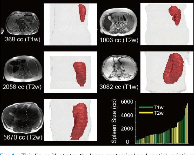
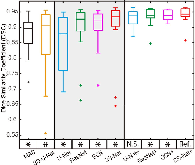
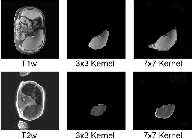
Abstract:The findings of splenomegaly, abnormal enlargement of the spleen, is a non-invasive clinical biomarker for liver and spleen disease. Automated segmentation methods are essential to efficiently quantify splenomegaly from clinically acquired abdominal magnetic resonance imaging (MRI) scans. However, the task is challenging due to (1) large anatomical and spatial variations of splenomegaly, (2) large inter- and intra-scan intensity variations on multi-modal MRI, and (3) limited numbers of labeled splenomegaly scans. In this paper, we propose the Splenomegaly Segmentation Network (SS-Net) to introduce the deep convolutional neural network (DCNN) approaches in multi-modal MRI splenomegaly segmentation. Large convolutional kernel layers were used to address the spatial and anatomical variations, while the conditional generative adversarial networks (GAN) were employed to leverage the segmentation performance of SS-Net in an end-to-end manner. A clinically acquired cohort containing both T1-weighted (T1w) and T2-weighted (T2w) MRI splenomegaly scans was used to train and evaluate the performance of multi-atlas segmentation (MAS), 2D DCNN networks, and a 3D DCNN network. From the experimental results, the DCNN methods achieved superior performance to the state-of-the-art MAS method. The proposed SS-Net method achieved the highest median and mean Dice scores among investigated baseline DCNN methods.
SynSeg-Net: Synthetic Segmentation Without Target Modality Ground Truth
Oct 15, 2018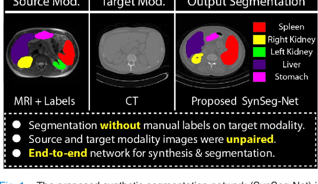
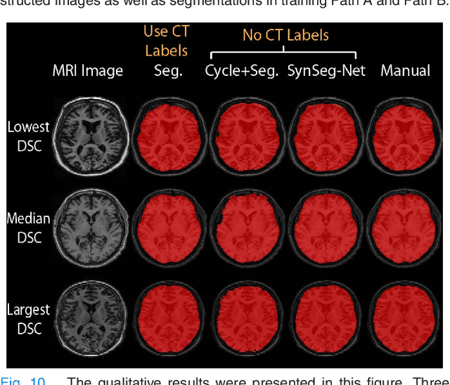
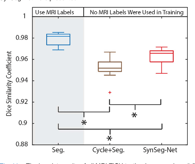

Abstract:A key limitation of deep convolutional neural networks (DCNN) based image segmentation methods is the lack of generalizability. Manually traced training images are typically required when segmenting organs in a new imaging modality or from distinct disease cohort. The manual efforts can be alleviated if the manually traced images in one imaging modality (e.g., MRI) are able to train a segmentation network for another imaging modality (e.g., CT). In this paper, we propose an end-to-end synthetic segmentation network (SynSeg-Net) to train a segmentation network for a target imaging modality without having manual labels. SynSeg-Net is trained by using (1) unpaired intensity images from source and target modalities, and (2) manual labels only from source modality. SynSeg-Net is enabled by the recent advances of cycle generative adversarial networks (CycleGAN) and DCNN. We evaluate the performance of the SynSeg-Net on two experiments: (1) MRI to CT splenomegaly synthetic segmentation for abdominal images, and (2) CT to MRI total intracranial volume synthetic segmentation (TICV) for brain images. The proposed end-to-end approach achieved superior performance to two stage methods. Moreover, the SynSeg-Net achieved comparable performance to the traditional segmentation network using target modality labels in certain scenarios. The source code of SynSeg-Net is publicly available (https://github.com/MASILab/SynSeg-Net).
Adversarial Synthesis Learning Enables Segmentation Without Target Modality Ground Truth
Dec 20, 2017


Abstract:A lack of generalizability is one key limitation of deep learning based segmentation. Typically, one manually labels new training images when segmenting organs in different imaging modalities or segmenting abnormal organs from distinct disease cohorts. The manual efforts can be alleviated if one is able to reuse manual labels from one modality (e.g., MRI) to train a segmentation network for a new modality (e.g., CT). Previously, two stage methods have been proposed to use cycle generative adversarial networks (CycleGAN) to synthesize training images for a target modality. Then, these efforts trained a segmentation network independently using synthetic images. However, these two independent stages did not use the complementary information between synthesis and segmentation. Herein, we proposed a novel end-to-end synthesis and segmentation network (EssNet) to achieve the unpaired MRI to CT image synthesis and CT splenomegaly segmentation simultaneously without using manual labels on CT. The end-to-end EssNet achieved significantly higher median Dice similarity coefficient (0.9188) than the two stages strategy (0.8801), and even higher than canonical multi-atlas segmentation (0.9125) and ResNet method (0.9107), which used the CT manual labels.
Splenomegaly Segmentation using Global Convolutional Kernels and Conditional Generative Adversarial Networks
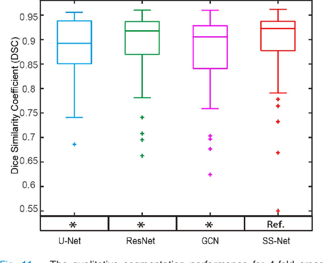
Dec 02, 2017Abstract:Spleen volume estimation using automated image segmentation technique may be used to detect splenomegaly (abnormally enlarged spleen) on Magnetic Resonance Imaging (MRI) scans. In recent years, Deep Convolutional Neural Networks (DCNN) segmentation methods have demonstrated advantages for abdominal organ segmentation. However, variations in both size and shape of the spleen on MRI images may result in large false positive and false negative labeling when deploying DCNN based methods. In this paper, we propose the Splenomegaly Segmentation Network (SSNet) to address spatial variations when segmenting extraordinarily large spleens. SSNet was designed based on the framework of image-to-image conditional generative adversarial networks (cGAN). Specifically, the Global Convolutional Network (GCN) was used as the generator to reduce false negatives, while the Markovian discriminator (PatchGAN) was used to alleviate false positives. A cohort of clinically acquired 3D MRI scans (both T1 weighted and T2 weighted) from patients with splenomegaly were used to train and test the networks. The experimental results demonstrated that a mean Dice coefficient of 0.9260 and a median Dice coefficient of 0.9262 using SSNet on independently tested MRI volumes of patients with splenomegaly.
 Add to Chrome
 Add to Firefox
 Add to Edge