Zunaira Rauf
Channel Boosted CNN-Transformer-based Multi-Level and Multi-Scale Nuclei Segmentation
Jul 27, 2024Abstract:Accurate nuclei segmentation is an essential foundation for various applications in computational pathology, including cancer diagnosis and treatment planning. Even slight variations in nuclei representations can significantly impact these downstream tasks. However, achieving accurate segmentation remains challenging due to factors like clustered nuclei, high intra-class variability in size and shape, resemblance to other cells, and color or contrast variations between nuclei and background. Despite the extensive utilization of Convolutional Neural Networks (CNNs) in medical image segmentation, they may have trouble capturing long-range dependencies crucial for accurate nuclei delineation. Transformers address this limitation but might miss essential low-level features. To overcome these limitations, we utilized CNN-Transformer-based techniques for nuclei segmentation in H&E stained histology images. In this work, we proposed two CNN-Transformer architectures, Nuclei Hybrid Vision Transformer (NucleiHVT) and Channel Boosted Nuclei Hybrid Vision Transformer (CB-NucleiHVT), that leverage the strengths of both CNNs and Transformers to effectively learn nuclei boundaries in multi-organ histology images. The first architecture, NucleiHVT is inspired by the UNet architecture and incorporates the dual attention mechanism to capture both multi-level and multi-scale context effectively. The CB-NucleiHVT network, on the other hand, utilizes the concept of channel boosting to learn diverse feature spaces, enhancing the model's ability to distinguish subtle variations in nuclei characteristics. Detailed evaluation of two medical image segmentation datasets shows that the proposed architectures outperform existing CNN-based, Transformer-based, and hybrid methods. The proposed networks demonstrated effective results both in terms of quantitative metrics, and qualitative visual assessment.
A Recent Survey of Vision Transformers for Medical Image Segmentation
Dec 01, 2023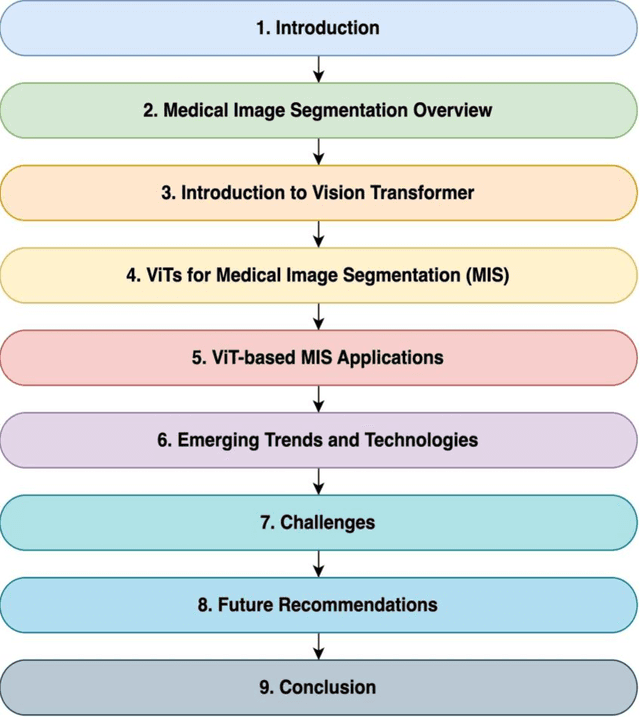
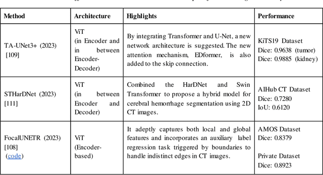

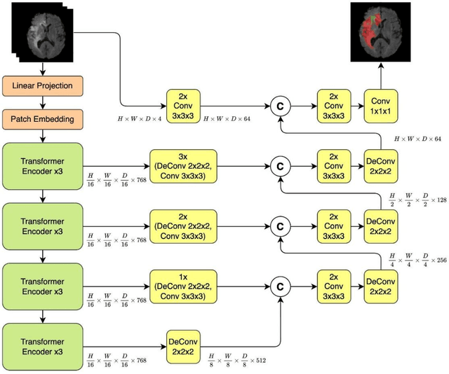
Abstract:Medical image segmentation plays a crucial role in various healthcare applications, enabling accurate diagnosis, treatment planning, and disease monitoring. In recent years, Vision Transformers (ViTs) have emerged as a promising technique for addressing the challenges in medical image segmentation. In medical images, structures are usually highly interconnected and globally distributed. ViTs utilize their multi-scale attention mechanism to model the long-range relationships in the images. However, they do lack image-related inductive bias and translational invariance, potentially impacting their performance. Recently, researchers have come up with various ViT-based approaches that incorporate CNNs in their architectures, known as Hybrid Vision Transformers (HVTs) to capture local correlation in addition to the global information in the images. This survey paper provides a detailed review of the recent advancements in ViTs and HVTs for medical image segmentation. Along with the categorization of ViT and HVT-based medical image segmentation approaches we also present a detailed overview of their real-time applications in several medical image modalities. This survey may serve as a valuable resource for researchers, healthcare practitioners, and students in understanding the state-of-the-art approaches for ViT-based medical image segmentation.
A survey of the Vision Transformers and its CNN-Transformer based Variants
May 25, 2023Abstract:Vision transformers have recently become popular as a possible alternative to convolutional neural networks (CNNs) for a variety of computer vision applications. These vision transformers due to their ability to focus on global relationships in images have large capacity, but may result in poor generalization as compared to CNNs. Very recently, the hybridization of convolution and self-attention mechanisms in vision transformers is gaining popularity due to their ability of exploiting both local and global image representations. These CNN-Transformer architectures also known as hybrid vision transformers have shown remarkable results for vision applications. Recently, due to the rapidly growing number of these hybrid vision transformers, there is a need for a taxonomy and explanation of these architectures. This survey presents a taxonomy of the recent vision transformer architectures, and more specifically that of the hybrid vision transformers. Additionally, the key features of each architecture such as the attention mechanisms, positional embeddings, multi-scale processing, and convolution are also discussed. This survey highlights the potential of hybrid vision transformers to achieve outstanding performance on a variety of computer vision tasks. Moreover, it also points towards the future directions of this rapidly evolving field.
CB-HVTNet: A channel-boosted hybrid vision transformer network for lymphocyte assessment in histopathological images
May 16, 2023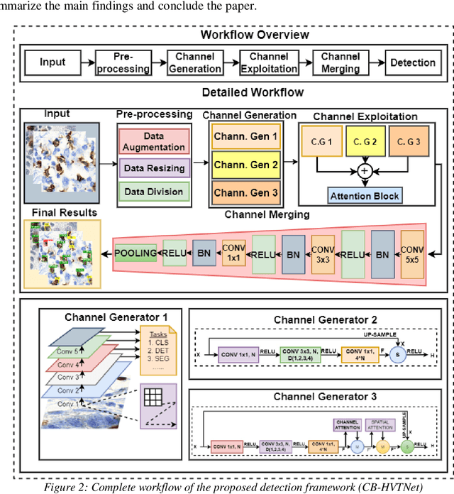

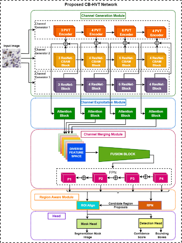

Abstract:Transformers, due to their ability to learn long range dependencies, have overcome the shortcomings of convolutional neural networks (CNNs) for global perspective learning. Therefore, they have gained the focus of researchers for several vision related tasks including medical diagnosis. However, their multi-head attention module only captures global level feature representations, which is insufficient for medical images. To address this issue, we propose a Channel Boosted Hybrid Vision Transformer (CB HVT) that uses transfer learning to generate boosted channels and employs both transformers and CNNs to analyse lymphocytes in histopathological images. The proposed CB HVT comprises five modules, including a channel generation module, channel exploitation module, channel merging module, region-aware module, and a detection and segmentation head, which work together to effectively identify lymphocytes. The channel generation module uses the idea of channel boosting through transfer learning to extract diverse channels from different auxiliary learners. In the CB HVT, these boosted channels are first concatenated and ranked using an attention mechanism in the channel exploitation module. A fusion block is then utilized in the channel merging module for a gradual and systematic merging of the diverse boosted channels to improve the network's learning representations. The CB HVT also employs a proposal network in its region aware module and a head to effectively identify objects, even in overlapping regions and with artifacts. We evaluated the proposed CB HVT on two publicly available datasets for lymphocyte assessment in histopathological images. The results show that CB HVT outperformed other state of the art detection models, and has good generalization ability, demonstrating its value as a tool for pathologists.
 Add to Chrome
 Add to Firefox
 Add to Edge