Wouter B. Veldhuis
Deep Group-wise Variational Diffeomorphic Image Registration
Oct 01, 2020


Abstract:Deep neural networks are increasingly used for pair-wise image registration. We propose to extend current learning-based image registration to allow simultaneous registration of multiple images. To achieve this, we build upon the pair-wise variational and diffeomorphic VoxelMorph approach and present a general mathematical framework that enables both registration of multiple images to their geodesic average and registration in which any of the available images can be used as a fixed image. In addition, we provide a likelihood based on normalized mutual information, a well-known image similarity metric in registration, between multiple images, and a prior that allows for explicit control over the viscous fluid energy to effectively regularize deformations. We trained and evaluated our approach using intra-patient registration of breast MRI and Thoracic 4DCT exams acquired over multiple time points. Comparison with Elastix and VoxelMorph demonstrates competitive quantitative performance of the proposed method in terms of image similarity and reference landmark distances at significantly faster registration.
Multimodal Learning for Cardiovascular Risk Prediction using EHR Data
Aug 27, 2020


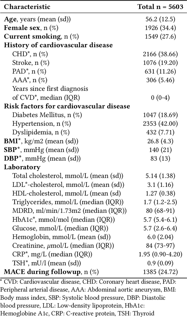
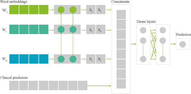
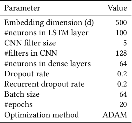
Abstract:Electronic health records (EHRs) contain structured and unstructured data of significant clinical and research value. Various machine learning approaches have been developed to employ information in EHRs for risk prediction. The majority of these attempts, however, focus on structured EHR fields and lose the vast amount of information in the unstructured texts. To exploit the potential information captured in EHRs, in this study we propose a multimodal recurrent neural network model for cardiovascular risk prediction that integrates both medical texts and structured clinical information. The proposed multimodal bidirectional long short-term memory (BiLSTM) model concatenates word embeddings to classical clinical predictors before applying them to a final fully connected neural network. In the experiments, we compare performance of different deep neural network (DNN) architectures including convolutional neural network and long short-term memory in scenarios of using clinical variables and chest X-ray radiology reports. Evaluated on a data set of real world patients with manifest vascular disease or at high-risk for cardiovascular disease, the proposed BiLSTM model demonstrates state-of-the-art performance and outperforms other DNN baseline architectures.
Liver segmentation and metastases detection in MR images using convolutional neural networks
Oct 15, 2019


Abstract:Primary tumors have a high likelihood of developing metastases in the liver and early detection of these metastases is crucial for patient outcome. We propose a method based on convolutional neural networks (CNN) to detect liver metastases. First, the liver was automatically segmented using the six phases of abdominal dynamic contrast enhanced (DCE) MR images. Next, DCE-MR and diffusion weighted (DW) MR images are used for metastases detection within the liver mask. The liver segmentations have a median Dice similarity coefficient of 0.95 compared with manual annotations. The metastases detection method has a sensitivity of 99.8% with a median of 2 false positives per image. The combination of the two MR sequences in a dual pathway network is proven valuable for the detection of liver metastases. In conclusion, a high quality liver segmentation can be obtained in which we can successfully detect liver metastases.
Motion correction of dynamic contrast enhanced MRI of the liver
Aug 22, 2019Abstract:Motion correction of dynamic contrast enhanced magnetic resonance images (DCE-MRI) is a challenging task, due to changes in image appearance. In this study a groupwise registration, using a principle component analysis (PCA) based metric,1 is evaluated for clinical DCE MRI of the liver. The groupwise registration transforms the images to a common space, rather than to a reference volume as conventional pairwise methods do, and computes the similarity metric on all volumes simultaneously. This groupwise registration method is compared to a pairwise approach using a mutual information metric. Clinical DCE MRI of the abdomen of eight patients were included. Per patient one lesion in the liver was manually segmented in all temporal images (N=16). The registered images were compared for accuracy, spatial and temporal smoothness after transformation, and lesion volume change. Compared to a pairwise method or no registration, groupwise registration provided better alignment. In our recently started clinical study groupwise registered clinical DCE MRI of the abdomen of nine patients were scored by three radiologists. Groupwise registration increased the assessed quality of alignment. The gain in reading time for the radiologist was estimated to vary from no difference to almost a minute. A slight increase in reader confidence was also observed. Registration had no added value for images with little motion. In conclusion, the groupwise registration of DCE MR images results in better alignment than achieved by pairwise registration, which is beneficial for clinical assessment.
 Add to Chrome
 Add to Firefox
 Add to Edge