Thomas Langø
Segmentation of Non-Small Cell Lung Carcinomas: Introducing DRU-Net and Multi-Lens Distortion
Jun 20, 2024
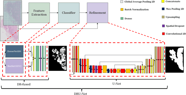
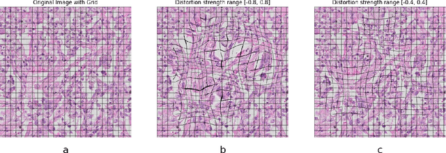

Abstract:Considering the increased workload in pathology laboratories today, automated tools such as artificial intelligence models can help pathologists with their tasks and ease the workload. In this paper, we are proposing a segmentation model (DRU-Net) that can provide a delineation of human non-small cell lung carcinomas and an augmentation method that can improve classification results. The proposed model is a fused combination of truncated pre-trained DenseNet201 and ResNet101V2 as a patch-wise classifier followed by a lightweight U-Net as a refinement model. We have used two datasets (Norwegian Lung Cancer Biobank and Haukeland University Hospital lung cancer cohort) to create our proposed model. The DRU-Net model achieves an average of 0.91 Dice similarity coefficient. The proposed spatial augmentation method (multi-lens distortion) improved the network performance by 3%. Our findings show that choosing image patches that specifically include regions of interest leads to better results for the patch-wise classifier compared to other sampling methods. The qualitative analysis showed that the DRU-Net model is generally successful in detecting the tumor. On the test set, some of the cases showed areas of false positive and false negative segmentation in the periphery, particularly in tumors with inflammatory and reactive changes.
AeroPath: An airway segmentation benchmark dataset with challenging pathology
Nov 02, 2023


Abstract:To improve the prognosis of patients suffering from pulmonary diseases, such as lung cancer, early diagnosis and treatment are crucial. The analysis of CT images is invaluable for diagnosis, whereas high quality segmentation of the airway tree are required for intervention planning and live guidance during bronchoscopy. Recently, the Multi-domain Airway Tree Modeling (ATM'22) challenge released a large dataset, both enabling training of deep-learning based models and bringing substantial improvement of the state-of-the-art for the airway segmentation task. However, the ATM'22 dataset includes few patients with severe pathologies affecting the airway tree anatomy. In this study, we introduce a new public benchmark dataset (AeroPath), consisting of 27 CT images from patients with pathologies ranging from emphysema to large tumors, with corresponding trachea and bronchi annotations. Second, we present a multiscale fusion design for automatic airway segmentation. Models were trained on the ATM'22 dataset, tested on the AeroPath dataset, and further evaluated against competitive open-source methods. The same performance metrics as used in the ATM'22 challenge were used to benchmark the different considered approaches. Lastly, an open web application is developed, to easily test the proposed model on new data. The results demonstrated that our proposed architecture predicted topologically correct segmentations for all the patients included in the AeroPath dataset. The proposed method is robust and able to handle various anomalies, down to at least the fifth airway generation. In addition, the AeroPath dataset, featuring patients with challenging pathologies, will contribute to development of new state-of-the-art methods. The AeroPath dataset and the web application are made openly available.
AI-Dentify: Deep learning for proximal caries detection on bitewing x-ray -- HUNT4 Oral Health Study
Sep 30, 2023Abstract:Background: Dental caries diagnosis requires the manual inspection of diagnostic bitewing images of the patient, followed by a visual inspection and probing of the identified dental pieces with potential lesions. Yet the use of artificial intelligence, and in particular deep-learning, has the potential to aid in the diagnosis by providing a quick and informative analysis of the bitewing images. Methods: A dataset of 13,887 bitewings from the HUNT4 Oral Health Study were annotated individually by six different experts, and used to train three different object detection deep-learning architectures: RetinaNet (ResNet50), YOLOv5 (M size), and EfficientDet (D0 and D1 sizes). A consensus dataset of 197 images, annotated jointly by the same six dentist, was used for evaluation. A five-fold cross validation scheme was used to evaluate the performance of the AI models. Results: the trained models show an increase in average precision and F1-score, and decrease of false negative rate, with respect to the dental clinicians. Out of the three architectures studied, YOLOv5 shows the largest improvement, reporting 0.647 mean average precision, 0.548 mean F1-score, and 0.149 mean false negative rate. Whereas the best annotators on each of these metrics reported 0.299, 0.495, and 0.164 respectively. Conclusion: Deep-learning models have shown the potential to assist dental professionals in the diagnosis of caries. Yet, the task remains challenging due to the artifacts natural to the bitewings.
Train smarter, not harder: learning deep abdominal CT registration on scarce data
Nov 30, 2022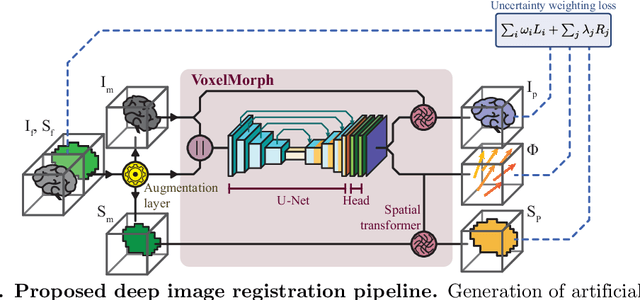
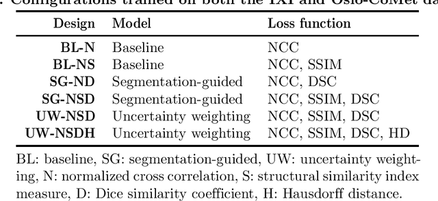


Abstract:Purpose: This study aims to explore training strategies to improve convolutional neural network-based image-to-image registration for abdominal imaging. Methods: Different training strategies, loss functions, and transfer learning schemes were considered. Furthermore, an augmentation layer which generates artificial training image pairs on-the-fly was proposed, in addition to a loss layer that enables dynamic loss weighting. Results: Guiding registration using segmentations in the training step proved beneficial for deep-learning-based image registration. Finetuning the pretrained model from the brain MRI dataset to the abdominal CT dataset further improved performance on the latter application, removing the need for a large dataset to yield satisfactory performance. Dynamic loss weighting also marginally improved performance, all without impacting inference runtime. Conclusion: Using simple concepts, we improved the performance of a commonly used deep image registration architecture, VoxelMorph. In future work, our framework, DDMR, should be validated on different datasets to further assess its value.
Teacher-Student Architecture for Mixed Supervised Lung Tumor Segmentation
Dec 21, 2021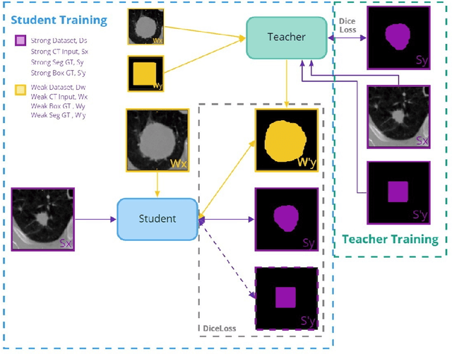
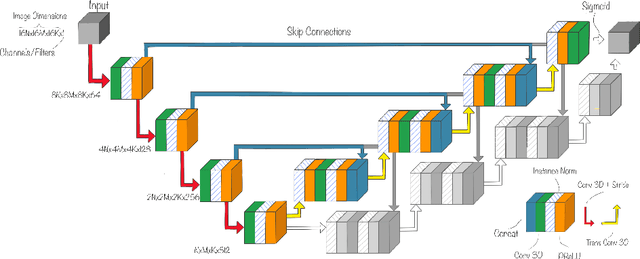


Abstract:Purpose: Automating tasks such as lung tumor localization and segmentation in radiological images can free valuable time for radiologists and other clinical personnel. Convolutional neural networks may be suited for such tasks, but require substantial amounts of labeled data to train. Obtaining labeled data is a challenge, especially in the medical domain. Methods: This paper investigates the use of a teacher-student design to utilize datasets with different types of supervision to train an automatic model performing pulmonary tumor segmentation on computed tomography images. The framework consists of two models: the student that performs end-to-end automatic tumor segmentation and the teacher that supplies the student additional pseudo-annotated data during training. Results: Using only a small proportion of semantically labeled data and a large number of bounding box annotated data, we achieved competitive performance using a teacher-student design. Models trained on larger amounts of semantic annotations did not perform better than those trained on teacher-annotated data. Conclusions: Our results demonstrate the potential of utilizing teacher-student designs to reduce the annotation load, as less supervised annotation schemes may be performed, without any real degradation in segmentation accuracy.
Mediastinal lymph nodes segmentation using 3D convolutional neural network ensembles and anatomical priors guiding
Feb 11, 2021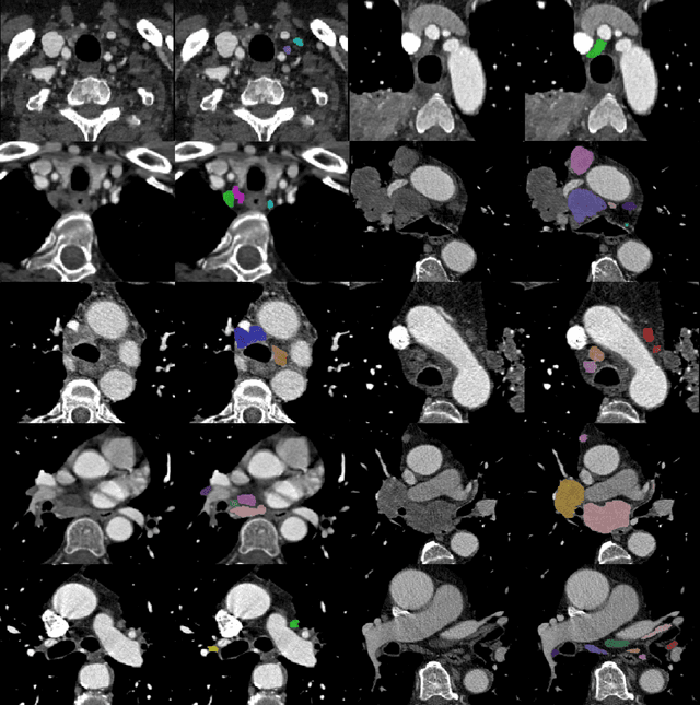
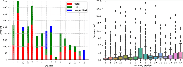

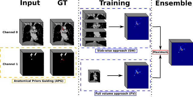
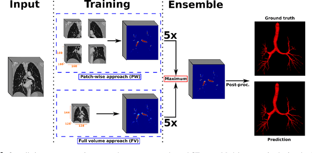
Abstract:As lung cancer evolves, the presence of enlarged and potentially malignant lymph nodes must be assessed to properly estimate disease progression and select the best treatment strategy. Following the clinical guidelines, estimation of short-axis diameter and mediastinum station are paramount for correct diagnosis. A method for accurate and automatic segmentation is hence decisive for quantitatively describing lymph nodes. In this study, the use of 3D convolutional neural networks, either through slab-wise schemes or the leveraging of downsampled entire volumes, is investigated. Furthermore, the potential impact from simple ensemble strategies is considered. As lymph nodes have similar attenuation values to nearby anatomical structures, we suggest using the knowledge of other organs as prior information to guide the segmentation task. To assess the segmentation and instance detection performances, a 5-fold cross-validation strategy was followed over a dataset of 120 contrast-enhanced CT volumes. For the 1178 lymph nodes with a short-axis diameter $\geq10$ mm, our best performing approach reached a patient-wise recall of 92%, a false positive per patient ratio of 5, and a segmentation overlap of 80.5%. The method performs similarly well across all stations. Fusing a slab-wise and a full volume approach within an ensemble scheme generated the best performances. The anatomical priors guiding strategy is promising, yet a larger set than four organs appears needed to generate an optimal benefit. A larger dataset is also mandatory, given the wide range of expressions a lymph node can exhibit (i.e., shape, location, and attenuation), and contrast uptake variations.
 Add to Chrome
 Add to Firefox
 Add to Edge