Segmentation of Non-Small Cell Lung Carcinomas: Introducing DRU-Net and Multi-Lens Distortion
Paper and Code
Jun 20, 2024


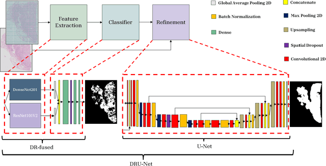
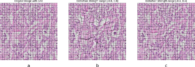
Considering the increased workload in pathology laboratories today, automated tools such as artificial intelligence models can help pathologists with their tasks and ease the workload. In this paper, we are proposing a segmentation model (DRU-Net) that can provide a delineation of human non-small cell lung carcinomas and an augmentation method that can improve classification results. The proposed model is a fused combination of truncated pre-trained DenseNet201 and ResNet101V2 as a patch-wise classifier followed by a lightweight U-Net as a refinement model. We have used two datasets (Norwegian Lung Cancer Biobank and Haukeland University Hospital lung cancer cohort) to create our proposed model. The DRU-Net model achieves an average of 0.91 Dice similarity coefficient. The proposed spatial augmentation method (multi-lens distortion) improved the network performance by 3%. Our findings show that choosing image patches that specifically include regions of interest leads to better results for the patch-wise classifier compared to other sampling methods. The qualitative analysis showed that the DRU-Net model is generally successful in detecting the tumor. On the test set, some of the cases showed areas of false positive and false negative segmentation in the periphery, particularly in tumors with inflammatory and reactive changes.
 Add to Chrome
 Add to Firefox
 Add to Edge