Sumeyra Demir Kanik
Prediction of Reaction Time and Vigilance Variability from Spatiospectral Features of Resting-State EEG in a Long Sustained Attention Task
Oct 21, 2019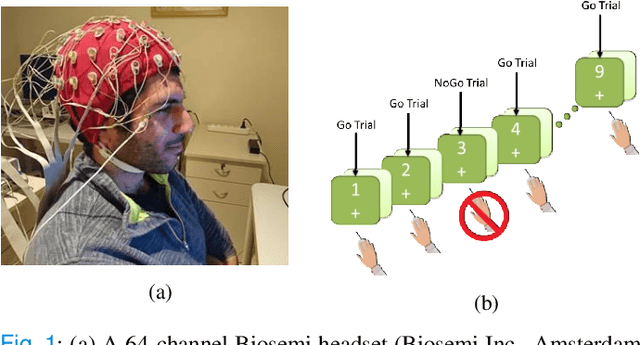


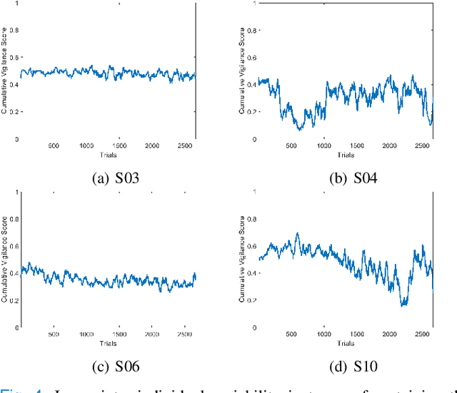
Abstract:Resting-state brain networks represent the intrinsic state of the brain during the majority of cognitive and sensorimotor tasks. However, no study has yet presented concise predictors of task-induced vigilance variability from spectrospatial features of the pre-task, resting-state electroencephalograms (EEG). We asked ten healthy volunteers (6 females, 4 males) to participate in 105-minute fixed-sequence-varying-duration sessions of sustained attention to response task (SART). A novel and adaptive vigilance scoring scheme was designed based on the performance and response time in consecutive trials, and demonstrated large inter-participant variability in terms of maintaining consistent tonic performance. Multiple linear regression using feature relevance analysis obtained significant predictors of the mean cumulative vigilance score (CVS), mean response time, and variabilities of these scores from the resting-state, band-power ratios of EEG signals, p<0.05. Single-layer neural networks trained with cross-validation also captured different associations for the beta sub-bands. Increase in the gamma (28-48 Hz) and upper beta ratios from the left central and temporal regions predicted slower reactions and more inconsistent vigilance as explained by the increased activation of default mode network (DMN) and differences between the high- and low-attention networks at temporal regions. Higher ratios of parietal alpha from the Brodmann's areas 18, 19, and 37 during the eyes-open states predicted slower responses but more consistent CVS and reactions associated with the superior ability in vigilance maintenance. The proposed framework and these findings on the most stable and significant attention predictors from the intrinsic EEG power ratios can be used to model attention variations during the calibration sessions of BCI applications and vigilance monitoring systems.
Dendritic Spine Shape Analysis: A Clustering Perspective
Jul 19, 2016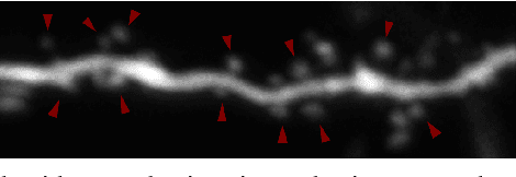

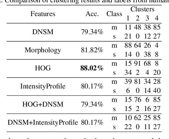
Abstract:Functional properties of neurons are strongly coupled with their morphology. Changes in neuronal activity alter morphological characteristics of dendritic spines. First step towards understanding the structure-function relationship is to group spines into main spine classes reported in the literature. Shape analysis of dendritic spines can help neuroscientists understand the underlying relationships. Due to unavailability of reliable automated tools, this analysis is currently performed manually which is a time-intensive and subjective task. Several studies on spine shape classification have been reported in the literature, however, there is an on-going debate on whether distinct spine shape classes exist or whether spines should be modeled through a continuum of shape variations. Another challenge is the subjectivity and bias that is introduced due to the supervised nature of classification approaches. In this paper, we aim to address these issues by presenting a clustering perspective. In this context, clustering may serve both confirmation of known patterns and discovery of new ones. We perform cluster analysis on two-photon microscopic images of spines using morphological, shape, and appearance based features and gain insights into the spine shape analysis problem. We use histogram of oriented gradients (HOG), disjunctive normal shape models (DNSM), morphological features, and intensity profile based features for cluster analysis. We use x-means to perform cluster analysis that selects the number of clusters automatically using the Bayesian information criterion (BIC). For all features, this analysis produces 4 clusters and we observe the formation of at least one cluster consisting of spines which are difficult to be assigned to a known class. This observation supports the argument of intermediate shape types.
 Add to Chrome
 Add to Firefox
 Add to Edge