Sonja Kirchhoff
Curriculum learning for annotation-efficient medical image analysis: scheduling data with prior knowledge and uncertainty
Jul 31, 2020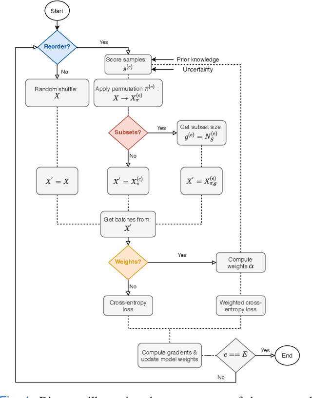
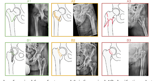
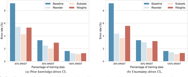
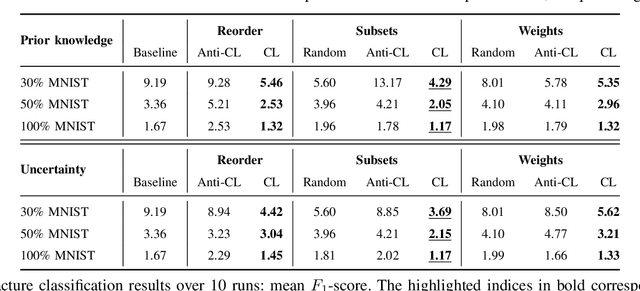
Abstract:Convolutional neural networks (CNNs) for multi-class classification require training on large, representative, and high quality annotated datasets. However, in the field of medical imaging, data and annotations are both difficult and expensive to acquire. Moreover, they frequently suffer from highly imbalanced distributions, and potentially noisy labels due to intra- or inter-expert disagreement. To deal with such challenges, we propose a unified curriculum learning framework to schedule the order and pace of the training samples presented to the optimizer. Our novel framework reunites three strategies consisting of individually weighting training samples, reordering the training set, or sampling subsets of data. The core of these strategies is a scoring function ranking the training samples according to either difficulty or uncertainty. We define the scoring function from domain-specific prior knowledge or by directly measuring the uncertainty in the predictions. We perform a variety of experiments with a clinical dataset for the multi-class classification of proximal femur fractures and the publicly available MNIST dataset. Our results show that the sequence and weight of the training samples play an important role in the optimization process of CNNs. Proximal femur fracture classification is improved up to the performance of experienced trauma surgeons. We further demonstrate the benefits of our unified curriculum learning method for three controlled and challenging digit recognition scenarios: with limited amounts of data, under class-imbalance, and in the presence of label noise.
Medical-based Deep Curriculum Learning for Improved Fracture Classification
Apr 01, 2020


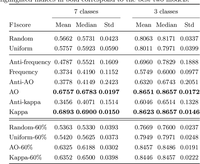
Abstract:Current deep-learning based methods do not easily integrate to clinical protocols, neither take full advantage of medical knowledge. In this work, we propose and compare several strategies relying on curriculum learning, to support the classification of proximal femur fracture from X-ray images, a challenging problem as reflected by existing intra- and inter-expert disagreement. Our strategies are derived from knowledge such as medical decision trees and inconsistencies in the annotations of multiple experts, which allows us to assign a degree of difficulty to each training sample. We demonstrate that if we start learning "easy" examples and move towards "hard", the model can reach a better performance, even with fewer data. The evaluation is performed on the classification of a clinical dataset of about 1000 X-ray images. Our results show that, compared to class-uniform and random strategies, the proposed medical knowledge-based curriculum, performs up to 15% better in terms of accuracy, achieving the performance of experienced trauma surgeons.
Towards an Interactive and Interpretable CAD System to Support Proximal Femur Fracture Classification
Feb 04, 2019


Abstract:Fractures of the proximal femur represent a critical entity in the western world, particularly with the growing elderly population. Such fractures result in high morbidity and mortality, reflecting a significant health and economic impact on our society. Different treatment strategies are recommended for different fracture types, with surgical treatment still being the gold standard in most of the cases. The success of the treatment and prognosis after surgery strongly depends on an accurate classification of the fracture among standard types, such as those defined by the AO system. However, the classification of fracture types based on x-ray images is difficult as confirmed by low intra- and inter-expert agreement rates of our in-house study and also in the previous literature. The presented work proposes a fully automatic computer-aided diagnosis (CAD) tool, based on current deep learning techniques, able to identify, localize and finally classify proximal femur fractures on x-rays images according to the AO classification. Results of our experimental evaluation show that the performance achieved by the proposed CAD tool is comparable to the average expert for the classification of x-ray images into types ''A'', ''B'' and ''normal'' (precision of 89%), while the performance is even superior when classifying fractures versus ''normal'' cases (precision of 94%). In addition, the integration of the proposed CAD tool into daily clinical routine is extensively discussed, towards improving the interface between humans and AI-powered machines in supporting medical decisions.
Weakly-Supervised Localization and Classification of Proximal Femur Fractures
Sep 27, 2018


Abstract:In this paper, we target the problem of fracture classification from clinical X-Ray images towards an automated Computer Aided Diagnosis (CAD) system. Although primarily dealing with an image classification problem, we argue that localizing the fracture in the image is crucial to make good class predictions. Therefore, we propose and thoroughly analyze several schemes for simultaneous fracture localization and classification. We show that using an auxiliary localization task, in general, improves the classification performance. Moreover, it is possible to avoid the need for additional localization annotations thanks to recent advancements in weakly-supervised deep learning approaches. Among such approaches, we investigate and adapt Spatial Transformers (ST), Self-Transfer Learning (STL), and localization from global pooling layers. We provide a detailed quantitative and qualitative validation on a dataset of 1347 femur fractures images and report high accuracy with regard to inter-expert correlation values reported in the literature. Our investigations show that i) lesion localization improves the classification outcome, ii) weakly-supervised methods improve baseline classification without any additional cost, iii) STL guides feature activations and boost performance. We plan to make both the dataset and code available.
 Add to Chrome
 Add to Firefox
 Add to Edge