Shuangzhi Lv
Deep learning to estimate the physical proportion of infected region of lung for COVID-19 pneumonia with CT image set
Jun 09, 2020


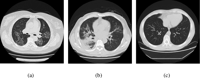
Abstract:Utilizing computed tomography (CT) images to quickly estimate the severity of cases with COVID-19 is one of the most straightforward and efficacious methods. Two tasks were studied in this present paper. One was to segment the mask of intact lung in case of pneumonia. Another was to generate the masks of regions infected by COVID-19. The masks of these two parts of images then were converted to corresponding volumes to calculate the physical proportion of infected region of lung. A total of 129 CT image set were herein collected and studied. The intrinsic Hounsfiled value of CT images was firstly utilized to generate the initial dirty version of labeled masks both for intact lung and infected regions. Then, the samples were carefully adjusted and improved by two professional radiologists to generate the final training set and test benchmark. Two deep learning models were evaluated: UNet and 2.5D UNet. For the segment of infected regions, a deep learning based classifier was followed to remove unrelated blur-edged regions that were wrongly segmented out such as air tube and blood vessel tissue etc. For the segmented masks of intact lung and infected regions, the best method could achieve 0.972 and 0.757 measure in mean Dice similarity coefficient on our test benchmark. As the overall proportion of infected region of lung, the final result showed 0.961 (Pearson's correlation coefficient) and 11.7% (mean absolute percent error). The instant proportion of infected regions of lung could be used as a visual evidence to assist clinical physician to determine the severity of the case. Furthermore, a quantified report of infected regions can help predict the prognosis for COVID-19 cases which were scanned periodically within the treatment cycle.
Deep Learning System to Screen Coronavirus Disease 2019 Pneumonia
Feb 21, 2020


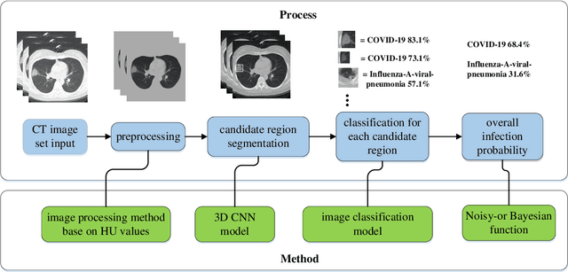
Abstract:We found that the real time reverse transcription-polymerase chain reaction (RT-PCR) detection of viral RNA from sputum or nasopharyngeal swab has a relatively low positive rate in the early stage to determine COVID-19 (named by the World Health Organization). The manifestations of computed tomography (CT) imaging of COVID-19 had their own characteristics, which are different from other types of viral pneumonia, such as Influenza-A viral pneumonia. Therefore, clinical doctors call for another early diagnostic criteria for this new type of pneumonia as soon as possible.This study aimed to establish an early screening model to distinguish COVID-19 pneumonia from Influenza-A viral pneumonia and healthy cases with pulmonary CT images using deep learning techniques. The candidate infection regions were first segmented out using a 3-dimensional deep learning model from pulmonary CT image set. These separated images were then categorized into COVID-19, Influenza-A viral pneumonia and irrelevant to infection groups, together with the corresponding confidence scores using a location-attention classification model. Finally the infection type and total confidence score of this CT case were calculated with Noisy-or Bayesian function.The experiments result of benchmark dataset showed that the overall accuracy was 86.7 % from the perspective of CT cases as a whole.The deep learning models established in this study were effective for the early screening of COVID-19 patients and demonstrated to be a promising supplementary diagnostic method for frontline clinical doctors.
 Add to Chrome
 Add to Firefox
 Add to Edge