Rupert Ecker
Fusion of Foundation and Vision Transformer Model Features for Dermatoscopic Image Classification
May 22, 2025Abstract:Accurate classification of skin lesions from dermatoscopic images is essential for diagnosis and treatment of skin cancer. In this study, we investigate the utility of a dermatology-specific foundation model, PanDerm, in comparison with two Vision Transformer (ViT) architectures (ViT base and Swin Transformer V2 base) for the task of skin lesion classification. Using frozen features extracted from PanDerm, we apply non-linear probing with three different classifiers, namely, multi-layer perceptron (MLP), XGBoost, and TabNet. For the ViT-based models, we perform full fine-tuning to optimize classification performance. Our experiments on the HAM10000 and MSKCC datasets demonstrate that the PanDerm-based MLP model performs comparably to the fine-tuned Swin transformer model, while fusion of PanDerm and Swin Transformer predictions leads to further performance improvements. Future work will explore additional foundation models, fine-tuning strategies, and advanced fusion techniques.
Automatic Foot Ulcer segmentation Using an Ensemble of Convolutional Neural Networks
Sep 03, 2021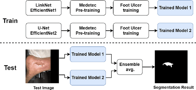
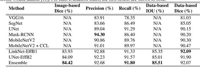
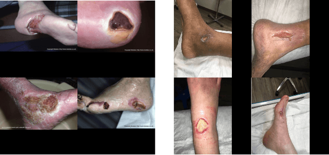

Abstract:Foot ulcer is a common complication of diabetes mellitus; it is associated with substantial morbidity and mortality and remains a major risk factor for lower leg amputation. Extracting accurate morphological features from the foot wounds is crucial for proper treatment. Although visual and manual inspection by medical professionals is the common approach to extract the features, this method is subjective and error-prone. Computer-mediated approaches are the alternative solutions to segment the lesions and extract related morphological features. Among various proposed computer-based approaches for image segmentation, deep learning-based methods and more specifically convolutional neural networks (CNN) have shown excellent performances for various image segmentation tasks including medical image segmentation. In this work, we proposed an ensemble approach based on two encoder-decoder-based CNN models, namely LinkNet and UNet, to perform foot ulcer segmentation. To deal with limited training samples, we used pre-trained weights (EfficientNetB1 for the LinkNet model and EfficientNetB2 for the UNet model) and further pre-training by the Medetec dataset. We also applied a number of morphological-based and colour-based augmentation techniques to train the models. We integrated five-fold cross-validation, test time augmentation and result fusion in our proposed ensemble approach to boost the segmentation performance. Applied on a publicly available foot ulcer segmentation dataset and the MICCAI 2021 Foot Ulcer Segmentation (FUSeg) Challenge, our method achieved state-of-the-art data-based Dice scores of 92.07% and 88.80%, respectively. Our developed method achieved the first rank in the FUSeg challenge leaderboard. The Dockerised guideline, inference codes and saved trained models are publicly available in the published GitHub repository: https://github.com/masih4/Foot_Ulcer_Segmentation
CryoNuSeg: A Dataset for Nuclei Instance Segmentation of Cryosectioned H&E-Stained Histological Images
Jan 02, 2021

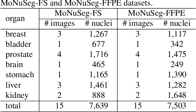
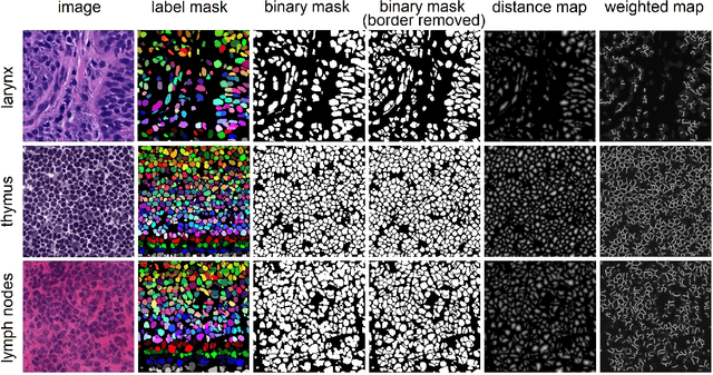
Abstract:Nuclei instance segmentation plays an important role in the analysis of Hematoxylin and Eosin (H&E)-stained images. While supervised deep learning (DL)-based approaches represent the state-of-the-art in automatic nuclei instance segmentation, annotated datasets are required to train these models. There are two main types of tissue processing protocols, namely formalin-fixed paraffin-embedded samples (FFPE) and frozen tissue samples (FS). Although FFPE-derived H&E stained tissue sections are the most widely used samples, H&E staining on frozen sections derived from FS samples is a relevant method in intra-operative surgical sessions as it can be performed fast. Due to differences in the protocols of these two types of samples, the derived images and in particular the nuclei appearance may be different in the acquired whole slide images. Analysis of FS-derived H&E stained images can be more challenging as rapid preparation, staining, and scanning of FS sections may lead to deterioration in image quality. In this paper, we introduce CryoNuSeg, the first fully annotated FS-derived cryosectioned and H&E-stained nuclei instance segmentation dataset. The dataset contains images from 10 human organs that were not exploited in other publicly available datasets, and is provided with three manual mark-ups to allow measuring intra-observer and inter-observer variability. Moreover, we investigate the effects of tissue fixation/embedding protocol (i.e., FS or FFPE) on the automatic nuclei instance segmentation performance of one of the state-of-the-art DL approaches. We also create a baseline segmentation benchmark for the dataset that can be used in future research. A step-by-step guide to generate the dataset as well as the full dataset and other detailed information are made available to fellow researchers at https://github.com/masih4/CryoNuSeg.
Pollen Grain Microscopic Image Classification Using an Ensemble of Fine-Tuned Deep Convolutional Neural Networks
Nov 15, 2020


Abstract:Pollen grain micrograph classification has multiple applications in medicine and biology. Automatic pollen grain image classification can alleviate the problems of manual categorisation such as subjectivity and time constraints. While a number of computer-based methods have been introduced in the literature to perform this task, classification performance needs to be improved for these methods to be useful in practice. In this paper, we present an ensemble approach for pollen grain microscopic image classification into four categories: Corylus Avellana well-developed pollen grain, Corylus Avellana anomalous pollen grain, Alnus well-developed pollen grain, and non-pollen (debris) instances. In our approach, we develop a classification strategy that is based on fusion of four state-of-the-art fine-tuned convolutional neural networks, namely EfficientNetB0, EfficientNetB1, EfficientNetB2 and SeResNeXt-50 deep models. These models are trained with images of three fixed sizes (224x224, 240x240, and 260x260 pixels) and their prediction probability vectors are then fused in an ensemble method to form a final classification vector for a given pollen grain image. Our proposed method is shown to yield excellent classification performance, obtaining an accuracy of of 94.48% and a weighted F1-score of 94.54% on the ICPR 2020 Pollen Grain Classification Challenge training dataset based on five-fold cross-validation. Evaluated on the test set of the challenge, our approach achieved a very competitive performance in comparison to the top ranked approaches with an accuracy and a weighted F1-score of 96.28% and 96.30%, respectively.
The Effects of Skin Lesion Segmentation on the Performance of Dermatoscopic Image Classification
Aug 28, 2020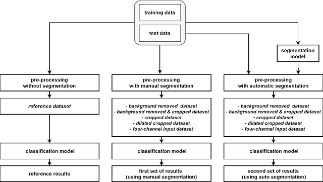
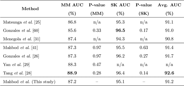
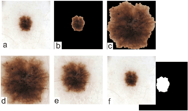
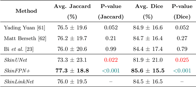
Abstract:Malignant melanoma (MM) is one of the deadliest types of skin cancer. Analysing dermatoscopic images plays an important role in the early detection of MM and other pigmented skin lesions. Among different computer-based methods, deep learning-based approaches and in particular convolutional neural networks have shown excellent classification and segmentation performances for dermatoscopic skin lesion images. These models can be trained end-to-end without requiring any hand-crafted features. However, the effect of using lesion segmentation information on classification performance has remained an open question. In this study, we explicitly investigated the impact of using skin lesion segmentation masks on the performance of dermatoscopic image classification. To do this, first, we developed a baseline classifier as the reference model without using any segmentation masks. Then, we used either manually or automatically created segmentation masks in both training and test phases in different scenarios and investigated the classification performances. Evaluated on the ISIC 2017 challenge dataset which contained two binary classification tasks (i.e. MM vs. all and seborrheic keratosis (SK) vs. all) and based on the derived area under the receiver operating characteristic curve scores, we observed four main outcomes. Our results show that 1) using segmentation masks did not significantly improve the MM classification performance in any scenario, 2) in one of the scenarios (using segmentation masks for dilated cropping), SK classification performance was significantly improved, 3) removing all background information by the segmentation masks significantly degraded the overall classification performance, and 4) in case of using the appropriate scenario (using segmentation for dilated cropping), there is no significant difference of using manually or automatically created segmentation masks.
Investigating and Exploiting Image Resolution for Transfer Learning-based Skin Lesion Classification
Jun 25, 2020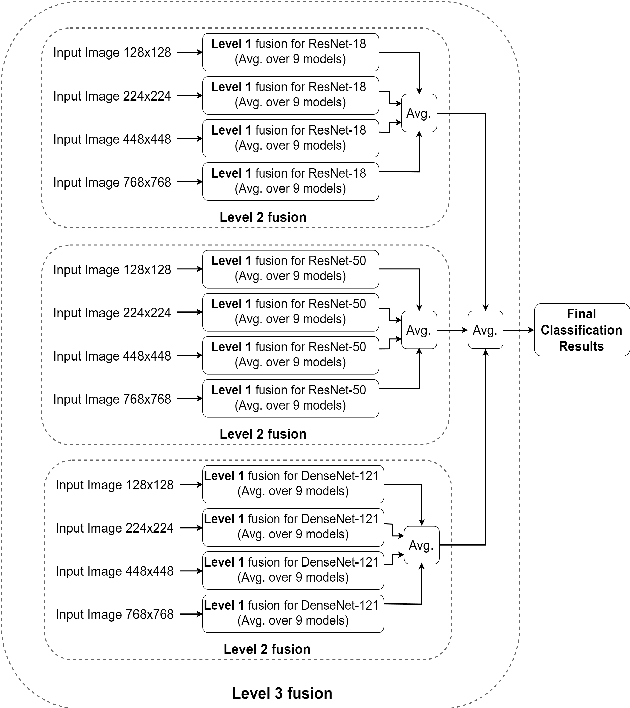

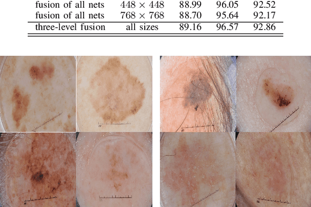

Abstract:Skin cancer is among the most common cancer types. Dermoscopic image analysis improves the diagnostic accuracy for detection of malignant melanoma and other pigmented skin lesions when compared to unaided visual inspection. Hence, computer-based methods to support medical experts in the diagnostic procedure are of great interest. Fine-tuning pre-trained convolutional neural networks (CNNs) has been shown to work well for skin lesion classification. Pre-trained CNNs are usually trained with natural images of a fixed image size which is typically significantly smaller than captured skin lesion images and consequently dermoscopic images are downsampled for fine-tuning. However, useful medical information may be lost during this transformation. In this paper, we explore the effect of input image size on skin lesion classification performance of fine-tuned CNNs. For this, we resize dermoscopic images to different resolutions, ranging from 64x64 to 768x768 pixels and investigate the resulting classification performance of three well-established CNNs, namely DenseNet-121, ResNet-18, and ResNet-50. Our results show that using very small images (of size 64x64 pixels) degrades the classification performance, while images of size 128x128 pixels and above support good performance with larger image sizes leading to slightly improved classification. We further propose a novel fusion approach based on a three-level ensemble strategy that exploits multiple fine-tuned networks trained with dermoscopic images at various sizes. When applied on the ISIC 2017 skin lesion classification challenge, our fusion approach yields an area under the receiver operating characteristic curve of 89.2% and 96.6% for melanoma classification and seborrheic keratosis classification, respectively, outperforming state-of-the-art algorithms.
Skin Lesion Classification Using Hybrid Deep Neural Networks
Feb 27, 2017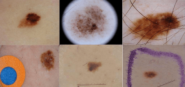
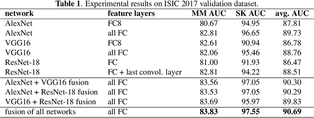

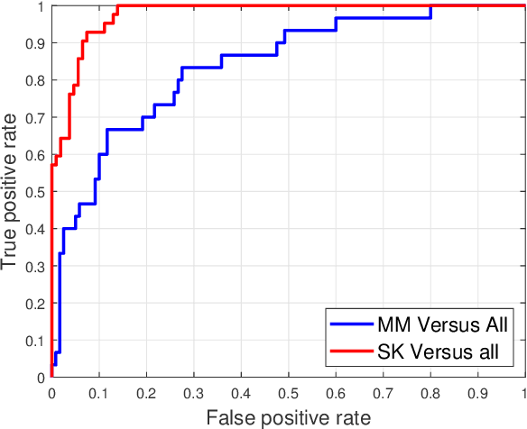
Abstract:Skin cancer is one of the major types of cancers and its incidence has been increasing over the past decades. Skin lesions can arise from various dermatologic disorders and can be classified to various types according to their texture, structure, color and other morphological features. The accuracy of diagnosis of skin lesions, specifically the discrimination of benign and malignant lesions, is paramount to ensure appropriate patient treatment. Machine learning-based classification approaches are among popular automatic methods for skin lesion classification. While there are many existing methods, convolutional neural networks (CNN) have shown to be superior over other classical machine learning methods for object detection and classification tasks. In this work, a fully automatic computerized method is proposed, which employs well established pre-trained convolutional neural networks and ensembles learning to classify skin lesions. We trained the networks using 2000 skin lesion images available from the ISIC 2017 challenge, which has three main categories and includes 374 melanoma, 254 seborrheic keratosis and 1372 benign nevi images. The trained classifier was then tested on 150 unlabeled images. The results, evaluated by the challenge organizer and based on the area under the receiver operating characteristic curve (AUC), were 84.8% and 93.6% for Melanoma and seborrheic keratosis binary classification problem, respectively. The proposed method achieved competitive results to experienced dermatologist. Further improvement and optimization of the proposed method with a larger training dataset could lead to a more precise, reliable and robust method for skin lesion classification.
 Add to Chrome
 Add to Firefox
 Add to Edge