Pengwei Wu
Coronary Atherosclerotic Plaque Characterization with Photon-counting CT: a Simulation-based Feasibility Study
Dec 04, 2023
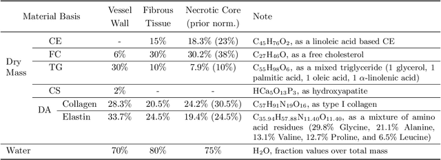
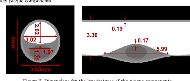

Abstract:Recent development of photon-counting CT (PCCT) brings great opportunities for plaque characterization with much-improved spatial resolution and spectral imaging capability. While existing coronary plaque PCCT imaging results are based on detectors made of CZT or CdTe materials, deep-silicon photon-counting detectors have unique performance characteristics and promise distinct imaging capabilities. In this work, we report a systematic simulation study of a deep-silicon PCCT scanner with a new clinically-relevant digital plaque phantom with realistic geometrical parameters and chemical compositions. This work investigates the effects of spatial resolution, noise, motion artifacts, radiation dose, and spectral characterization. Our simulation results suggest that the deep-silicon PCCT design provides adequate spatial resolution for visualizing a necrotic core and quantitation of key plaque features. Advanced denoising techniques and aggressive bowtie filter designs can keep image noise to acceptable levels at this resolution while keeping radiation dose comparable to that of a conventional CT scan. The ultrahigh resolution of PCCT also means an elevated sensitivity to motion artifacts. It is found that a tolerance of less than 0.4 mm residual movement range requires the application of accurate motion correction methods for best plaque imaging quality with PCCT.
Using Uncertainty in Deep Learning Reconstruction for Cone-Beam CT of the Brain
Aug 20, 2021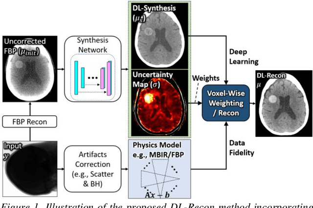
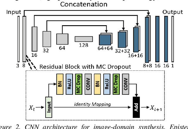
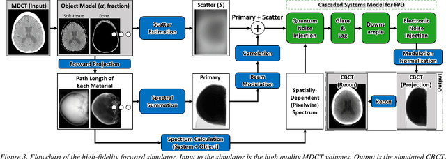
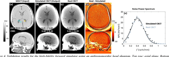
Abstract:Contrast resolution beyond the limits of conventional cone-beam CT (CBCT) systems is essential to high-quality imaging of the brain. We present a deep learning reconstruction method (dubbed DL-Recon) that integrates physically principled reconstruction models with DL-based image synthesis based on the statistical uncertainty in the synthesis image. A synthesis network was developed to generate a synthesized CBCT image (DL-Synthesis) from an uncorrected filtered back-projection (FBP) image. To improve generalizability (including accurate representation of lesions not seen in training), voxel-wise epistemic uncertainty of DL-Synthesis was computed using a Bayesian inference technique (Monte-Carlo dropout). In regions of high uncertainty, the DL-Recon method incorporates information from a physics-based reconstruction model and artifact-corrected projection data. Two forms of the DL-Recon method are proposed: (i) image-domain fusion of DL-Synthesis and FBP (DL-FBP) weighted by DL uncertainty; and (ii) a model-based iterative image reconstruction (MBIR) optimization using DL-Synthesis to compute a spatially varying regularization term based on DL uncertainty (DL-MBIR). The error in DL-Synthesis images was correlated with the uncertainty in the synthesis estimate. Compared to FBP and PWLS, the DL-Recon methods (both DL-FBP and DL-MBIR) showed ~50% reduction in noise (at matched spatial resolution) and ~40-70% improvement in image uniformity. Conventional DL-Synthesis alone exhibited ~10-60% under-estimation of lesion contrast and ~5-40% reduction in lesion segmentation accuracy (Dice coefficient) in simulated and real brain lesions, suggesting a lack of reliability / generalizability for structures unseen in the training data. DL-FBP and DL-MBIR improved the accuracy of reconstruction by directly incorporating information from the measurements in regions of high uncertainty.
 Add to Chrome
 Add to Firefox
 Add to Edge