Oleg Rogov
OrtSAE: Orthogonal Sparse Autoencoders Uncover Atomic Features
Sep 26, 2025Abstract:Sparse autoencoders (SAEs) are a technique for sparse decomposition of neural network activations into human-interpretable features. However, current SAEs suffer from feature absorption, where specialized features capture instances of general features creating representation holes, and feature composition, where independent features merge into composite representations. In this work, we introduce Orthogonal SAE (OrtSAE), a novel approach aimed to mitigate these issues by enforcing orthogonality between the learned features. By implementing a new training procedure that penalizes high pairwise cosine similarity between SAE features, OrtSAE promotes the development of disentangled features while scaling linearly with the SAE size, avoiding significant computational overhead. We train OrtSAE across different models and layers and compare it with other methods. We find that OrtSAE discovers 9% more distinct features, reduces feature absorption (by 65%) and composition (by 15%), improves performance on spurious correlation removal (+6%), and achieves on-par performance for other downstream tasks compared to traditional SAEs.
A Data-Centric Framework for Addressing Phonetic and Prosodic Challenges in Russian Speech Generative Models
Jul 17, 2025Abstract:Russian speech synthesis presents distinctive challenges, including vowel reduction, consonant devoicing, variable stress patterns, homograph ambiguity, and unnatural intonation. This paper introduces Balalaika, a novel dataset comprising more than 2,000 hours of studio-quality Russian speech with comprehensive textual annotations, including punctuation and stress markings. Experimental results show that models trained on Balalaika significantly outperform those trained on existing datasets in both speech synthesis and enhancement tasks. We detail the dataset construction pipeline, annotation methodology, and results of comparative evaluations.
Boundary-Aware Vision Transformer for Angiography Vascular Network Segmentation
Jun 15, 2025Abstract:Accurate segmentation of vascular structures in coronary angiography remains a core challenge in medical image analysis due to the complexity of elongated, thin, and low-contrast vessels. Classical convolutional neural networks (CNNs) often fail to preserve topological continuity, while recent Vision Transformer (ViT)-based models, although strong in global context modeling, lack precise boundary awareness. In this work, we introduce BAVT, a Boundary-Aware Vision Transformer, a ViT-based architecture enhanced with an edge-aware loss that explicitly guides the segmentation toward fine-grained vascular boundaries. Unlike hybrid transformer-CNN models, BAVT retains a minimal, scalable structure that is fully compatible with large-scale vision foundation model (VFM) pretraining. We validate our approach on the DCA-1 coronary angiography dataset, where BAVT achieves superior performance across medical image segmentation metrics outperforming both CNN and hybrid baselines. These results demonstrate the effectiveness of combining plain ViT encoders with boundary-aware supervision for clinical-grade vascular segmentation.
Spread them Apart: Towards Robust Watermarking of Generated Content
Feb 11, 2025Abstract:Generative models that can produce realistic images have improved significantly in recent years. The quality of the generated content has increased drastically, so sometimes it is very difficult to distinguish between the real images and the generated ones. Such an improvement comes at a price of ethical concerns about the usage of the generative models: the users of generative models can improperly claim ownership of the generated content protected by a license. In this paper, we propose an approach to embed watermarks into the generated content to allow future detection of the generated content and identification of the user who generated it. The watermark is embedded during the inference of the model, so the proposed approach does not require the retraining of the latter. We prove that watermarks embedded are guaranteed to be robust against additive perturbations of a bounded magnitude. We apply our method to watermark diffusion models and show that it matches state-of-the-art watermarking schemes in terms of robustness to different types of synthetic watermark removal attacks.
Suppressing Modulation Instability with Reinforcement Learning
Apr 05, 2024Abstract:Modulation instability is a phenomenon of spontaneous pattern formation in nonlinear media, oftentimes leading to an unpredictable behaviour and a degradation of a signal of interest. We propose an approach based on reinforcement learning to suppress the unstable modes by optimizing the parameters for the time modulation of the potential in the nonlinear system. We test our approach in 1D and 2D cases and propose a new class of physically-meaningful reward functions to guarantee tamed instability.
Probabilistically Robust Watermarking of Neural Networks
Jan 16, 2024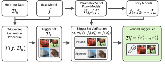
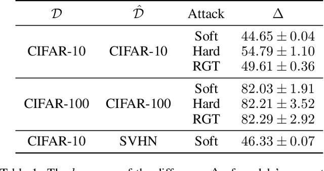
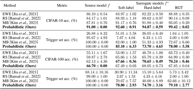
Abstract:As deep learning (DL) models are widely and effectively used in Machine Learning as a Service (MLaaS) platforms, there is a rapidly growing interest in DL watermarking techniques that can be used to confirm the ownership of a particular model. Unfortunately, these methods usually produce watermarks susceptible to model stealing attacks. In our research, we introduce a novel trigger set-based watermarking approach that demonstrates resilience against functionality stealing attacks, particularly those involving extraction and distillation. Our approach does not require additional model training and can be applied to any model architecture. The key idea of our method is to compute the trigger set, which is transferable between the source model and the set of proxy models with a high probability. In our experimental study, we show that if the probability of the set being transferable is reasonably high, it can be effectively used for ownership verification of the stolen model. We evaluate our method on multiple benchmarks and show that our approach outperforms current state-of-the-art watermarking techniques in all considered experimental setups.
Learnable Hollow Kernels for Anatomical Segmentation
Jul 09, 2020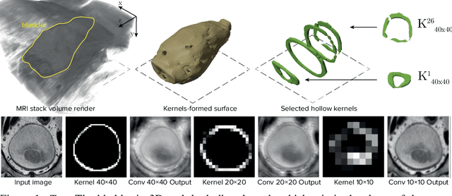

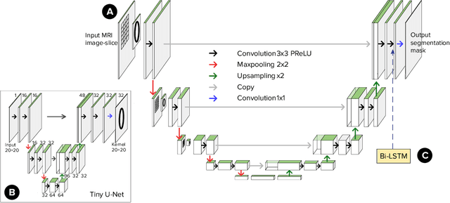
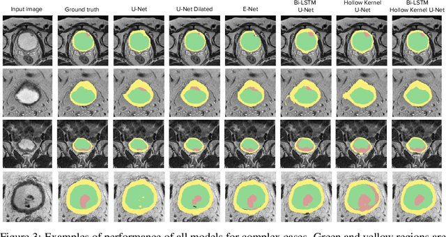
Abstract:Segmentation of certain hollow organs, such as the bladder, is especially hard to automate due to their complex geometry, vague intensity gradients in the soft tissues, and a tedious manual process of the data annotation routine. Yet, accurate localization of the walls and the cancer regions in the radiologic images of such organs is an essential step in oncology. To address this issue, we propose a new class of hollow kernels that learn to 'mimic' the contours of the segmented organ, effectively replicating its shape and structural complexity. We train a series of the U-Net-like neural networks using the proposed kernels and demonstrate the superiority of the idea in various spatio-temporal convolution scenarios. Specifically, the dilated hollow-kernel architecture outperforms state-of-the-art spatial segmentation models, whereas the addition of temporal blocks with, e.g., Bi-LSTM, establishes a new multi-class baseline for the bladder segmentation challenge. Our spatio-temporal model based on the hollow kernels reaches the mean dice scores of 0.936, 0.736, and 0.712 for the bladder's inner wall, the outer wall, and the tumor regions, respectively. The results pave the way towards other domain-specific deep learning applications where the shape of the segmented object could be used to form a proper convolution kernel for boosting the segmentation outcome.
Deep Negative Volume Segmentation
Jun 22, 2020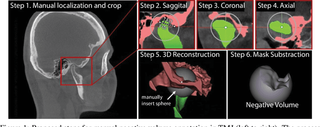

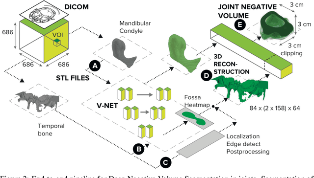
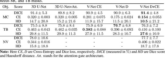
Abstract:Clinical examination of three-dimensional image data of compound anatomical objects, such as complex joints, remains a tedious process, demanding the time and the expertise of physicians. For instance, automation of the segmentation task of the TMJ (temporomandibular joint) has been hindered by its compound three-dimensional shape, multiple overlaid textures, an abundance of surrounding irregularities in the skull, and a virtually omnidirectional range of the jaw's motion - all of which extend the manual annotation process to more than an hour per patient. To address the challenge, we invent a new angle to the 3D segmentation task: namely, we propose to segment empty spaces between all the tissues surrounding the object - the so-called negative volume segmentation. Our approach is an end-to-end pipeline that comprises a V-Net for bone segmentation, a 3D volume construction by inflation of the reconstructed bone head in all directions along the normal vector to its mesh faces. Eventually confined within the skull bones, the inflated surface occupies the entire "negative" space in the joint, effectively providing a geometrical/topological metric of the joint's health. We validate the idea on the CT scans in a 50-patient dataset, annotated by experts in maxillofacial medicine, quantitatively compare the asymmetry given the left and the right negative volumes, and automate the entire framework for clinical adoption.
 Add to Chrome
 Add to Firefox
 Add to Edge