Mojtaba Masoudi
Computer Aided Detection for Pulmonary Embolism Challenge (CAD-PE)
Mar 30, 2020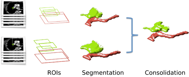
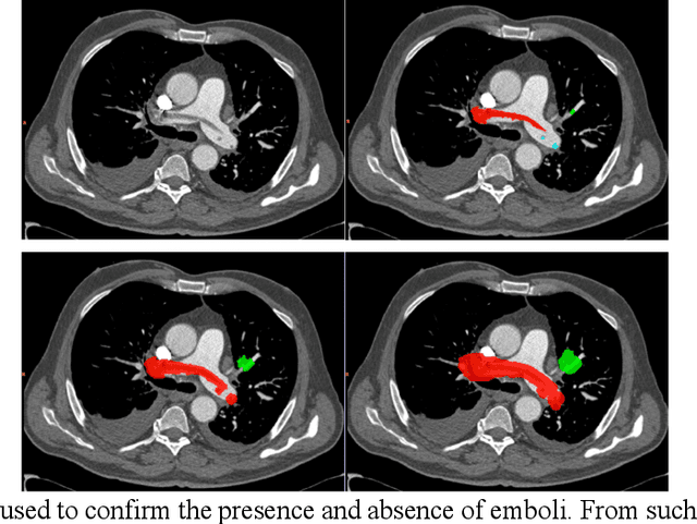
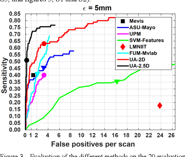
Abstract:Rationale: Computer aided detection (CAD) algorithms for Pulmonary Embolism (PE) algorithms have been shown to increase radiologists' sensitivity with a small increase in specificity. However, CAD for PE has not been adopted into clinical practice, likely because of the high number of false positives current CAD software produces. Objective: To generate a database of annotated computed tomography pulmonary angiographies, use it to compare the sensitivity and false positive rate of current algorithms and to develop new methods that improve such metrics. Methods: 91 Computed tomography pulmonary angiography scans were annotated by at least one radiologist by segmenting all pulmonary emboli visible on the study. 20 annotated CTPAs were open to the public in the form of a medical image analysis challenge. 20 more were kept for evaluation purposes. 51 were made available post-challenge. 8 submissions, 6 of them novel, were evaluated on the 20 evaluation CTPAs. Performance was measured as per embolus sensitivity vs. false positives per scan curve. Results: The best algorithms achieved a per-embolus sensitivity of 75% at 2 false positives per scan (fps) or of 70% at 1 fps, outperforming the state of the art. Deep learning approaches outperformed traditional machine learning ones, and their performance improved with the number of training cases. Significance: Through this work and challenge we have improved the state-of-the art of computer aided detection algorithms for pulmonary embolism. An open database and an evaluation benchmark for such algorithms have been generated, easing the development of further improvements. Implications on clinical practice will need further research.
Microaneurysm Detection in Fundus Images Using a Two-step Convolutional Neural Networks
Jul 08, 2018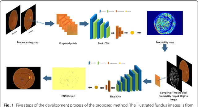
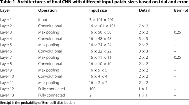
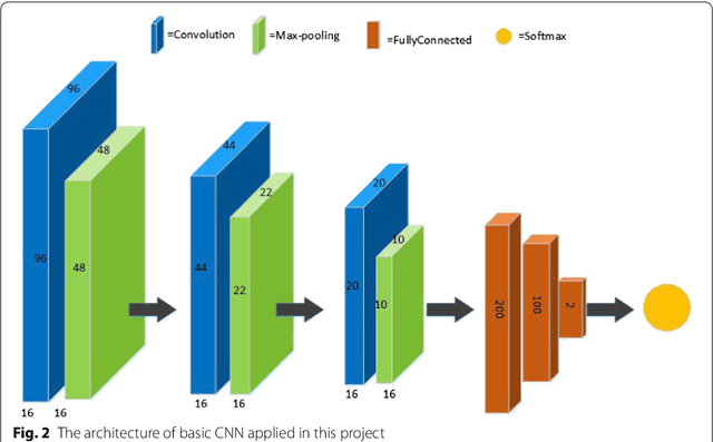
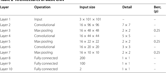
Abstract:Diabetic Retinopathy (DR) is a prominent cause of blindness in the world. The early treatment of DR can be conducted from detection of microaneurysms (MAs) which appears as reddish spots in retinal images. An automated microaneurysm detection can be a helpful system for ophthalmologists. In this paper, deep learning, in particular convolutional neural network (CNN), is used as a powerful tool to efficiently detect MAs from fundus images. In our method a new technique is used to utilise a two-stage training process which results in an accurate detection, while decreasing computational complexity in comparison with previous works. To validate our proposed method, an experiment is conducted using Keras library to implement our proposed CNN on two standard publicly available datasets. Our results show a promising sensitivity value of about 0.8 at the average number of false positive per image greater than 6 which is a competitive value with the state-of-the-art approaches.
A dataset for Computer-Aided Detection of Pulmonary Embolism in CTA images
Jul 05, 2017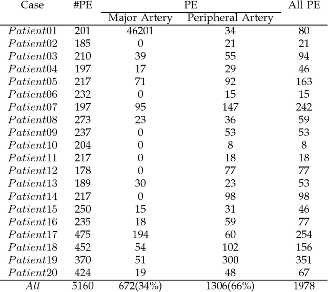
Abstract:Todays, researchers in the field of Pulmonary Embolism (PE) analysis need to use a publicly available dataset to assess and compare their methods. Different systems have been designed for the detection of pulmonary embolism (PE), but none of them have used any public datasets. All papers have used their own private dataset. In order to fill this gap, we have collected 5160 slices of computed tomography angiography (CTA) images acquired from 20 patients, and after labeling the image by experts in this field, we provided a reliable dataset which is now publicly available. In some situation, PE detection can be difficult, for example when it occurs in the peripheral branches or when patients have pulmonary diseases (such as parenchymal disease). Therefore, the efficiency of CAD systems highly depends on the dataset. In the given dataset, 66% of PE are located in peripheral branches, and different pulmonary diseases are also included.
 Add to Chrome
 Add to Firefox
 Add to Edge