Markus Axer
From Fibers to Cells: Fourier-Based Registration Enables Virtual Cresyl Violet Staining From 3D Polarized Light Imaging
May 16, 2025Abstract:Comprehensive assessment of the various aspects of the brain's microstructure requires the use of complementary imaging techniques. This includes measuring the spatial distribution of cell bodies (cytoarchitecture) and nerve fibers (myeloarchitecture). The gold standard for cytoarchitectonic analysis is light microscopic imaging of cell-body stained tissue sections. To reveal the 3D orientations of nerve fibers, 3D Polarized Light Imaging (3D-PLI) has been introduced as a reliable technique providing a resolution in the micrometer range while allowing processing of series of complete brain sections. 3D-PLI acquisition is label-free and allows subsequent staining of sections after measurement. By post-staining for cell bodies, a direct link between fiber- and cytoarchitecture can potentially be established within the same section. However, inevitable distortions introduced during the staining process make a nonlinear and cross-modal registration necessary in order to study the detailed relationships between cells and fibers in the images. In addition, the complexity of processing histological sections for post-staining only allows for a limited number of samples. In this work, we take advantage of deep learning methods for image-to-image translation to generate a virtual staining of 3D-PLI that is spatially aligned at the cellular level. In a supervised setting, we build on a unique dataset of brain sections, to which Cresyl violet staining has been applied after 3D-PLI measurement. To ensure high correspondence between both modalities, we address the misalignment of training data using Fourier-based registration methods. In this way, registration can be efficiently calculated during training for local image patches of target and predicted staining. We demonstrate that the proposed method enables prediction of a Cresyl violet staining from 3D-PLI, matching individual cell instances.
Analyzing Regional Organization of the Human Hippocampus in 3D-PLI Using Contrastive Learning and Geometric Unfolding
Feb 27, 2024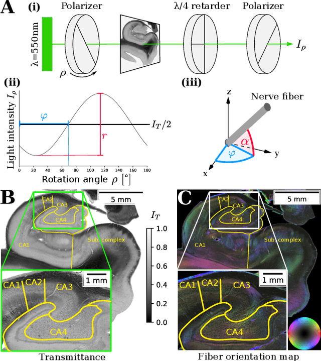
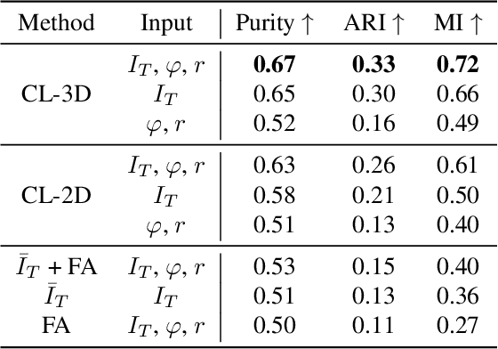
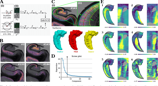
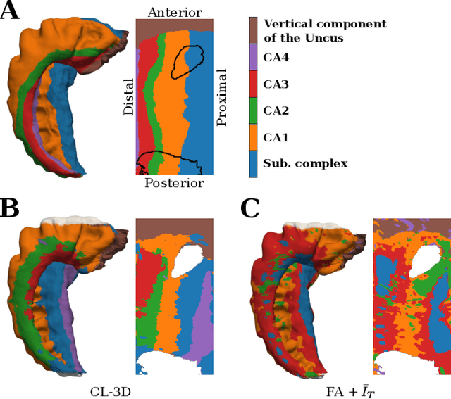
Abstract:Understanding the cortical organization of the human brain requires interpretable descriptors for distinct structural and functional imaging data. 3D polarized light imaging (3D-PLI) is an imaging modality for visualizing fiber architecture in postmortem brains with high resolution that also captures the presence of cell bodies, for example, to identify hippocampal subfields. The rich texture in 3D-PLI images, however, makes this modality particularly difficult to analyze and best practices for characterizing architectonic patterns still need to be established. In this work, we demonstrate a novel method to analyze the regional organization of the human hippocampus in 3D-PLI by combining recent advances in unfolding methods with deep texture features obtained using a self-supervised contrastive learning approach. We identify clusters in the representations that correspond well with classical descriptions of hippocampal subfields, lending validity to the developed methodology.
Self-Supervised Representation Learning for Nerve Fiber Distribution Patterns in 3D-PLI
Jan 30, 2024Abstract:A comprehensive understanding of the organizational principles in the human brain requires, among other factors, well-quantifiable descriptors of nerve fiber architecture. Three-dimensional polarized light imaging (3D-PLI) is a microscopic imaging technique that enables insights into the fine-grained organization of myelinated nerve fibers with high resolution. Descriptors characterizing the fiber architecture observed in 3D-PLI would enable downstream analysis tasks such as multimodal correlation studies, clustering, and mapping. However, best practices for observer-independent characterization of fiber architecture in 3D-PLI are not yet available. To this end, we propose the application of a fully data-driven approach to characterize nerve fiber architecture in 3D-PLI images using self-supervised representation learning. We introduce a 3D-Context Contrastive Learning (CL-3D) objective that utilizes the spatial neighborhood of texture examples across histological brain sections of a 3D reconstructed volume to sample positive pairs for contrastive learning. We combine this sampling strategy with specifically designed image augmentations to gain robustness to typical variations in 3D-PLI parameter maps. The approach is demonstrated for the 3D reconstructed occipital lobe of a vervet monkey brain. We show that extracted features are highly sensitive to different configurations of nerve fibers, yet robust to variations between consecutive brain sections arising from histological processing. We demonstrate their practical applicability for retrieving clusters of homogeneous fiber architecture and performing data mining for interactively selected templates of specific components of fiber architecture such as U-fibers.
GORDA: Graph-based ORientation Distribution Analysis of SLI scatterometry Patterns of Nerve Fibres
Apr 12, 2022


Abstract:Scattered Light Imaging (SLI) is a novel approach for microscopically revealing the fibre architecture of unstained brain sections. The measurements are obtained by illuminating brain sections from different angles and measuring the transmitted (scattered) light under normal incidence. The evaluation of scattering profiles commonly relies on a peak picking technique and feature extraction from the peaks, which allows quantitative determination of parallel and crossing in-plane nerve fibre directions for each image pixel. However, the estimation of the 3D orientation of the fibres cannot be assessed with the traditional methodology. We propose an unsupervised learning approach using spherical convolutions for estimating the 3D orientation of neural fibres, resulting in a more detailed interpretation of the fibre orientation distributions in the brain.
Towards ultra-high resolution 3D reconstruction of a whole rat brain from 3D-PLI data
Jul 29, 2018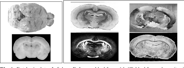

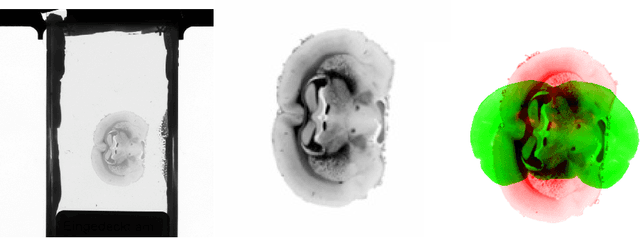

Abstract:3D reconstruction of the fiber connectivity of the rat brain at microscopic scale enables gaining detailed insight about the complex structural organization of the brain. We introduce a new method for registration and 3D reconstruction of high- and ultra-high resolution (64 $\mu$m and 1.3 $\mu$m pixel size) histological images of a Wistar rat brain acquired by 3D polarized light imaging (3D-PLI). Our method exploits multi-scale and multi-modal 3D-PLI data up to cellular resolution. We propose a new feature transform-based similarity measure and a weighted regularization scheme for accurate and robust non-rigid registration. To transform the 1.3 $\mu$m ultra-high resolution data to the reference blockface images a feature-based registration method followed by a non-rigid registration is proposed. Our approach has been successfully applied to 278 histological sections of a rat brain and the performance has been quantitatively evaluated using manually placed landmarks by an expert.
 Add to Chrome
 Add to Firefox
 Add to Edge