Kwame S. Kutten
3D Normal Coordinate Systems for Cortical Areas
Aug 07, 2018

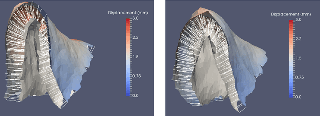
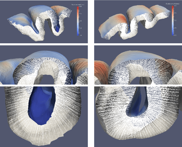
Abstract:A surface-based diffeomorphic algorithm to generate 3D coordinate grids in the cortical ribbon is described. In the grid, normal coordinate lines are generated by the diffeomorphic evolution from the grey/white (inner) surface to the grey/csf (outer) surface. Specifically, the cortical ribbon is described by two triangulated surfaces with open boundaries. Conceptually, the inner surface sits on top of the white matter structure and the outer on top of the gray matter. It is assumed that the cortical ribbon consists of cortical columns which are orthogonal to the white matter surface. This might be viewed as a consequence of the development of the columns in the embryo. It is also assumed that the columns are orthogonal to the outer surface so that the resultant vector field is orthogonal to the evolving surface. Then the distance of the normal lines from the vector field such that the inner surface evolves diffeomorphically towards the outer one can be construed as a measure of thickness. Applications are described for the auditory cortices in human adults and cats with normal hearing or hearing loss. The approach offers great potential for cortical morphometry.
A Large Deformation Diffeomorphic Approach to Registration of CLARITY Images via Mutual Information
Aug 11, 2017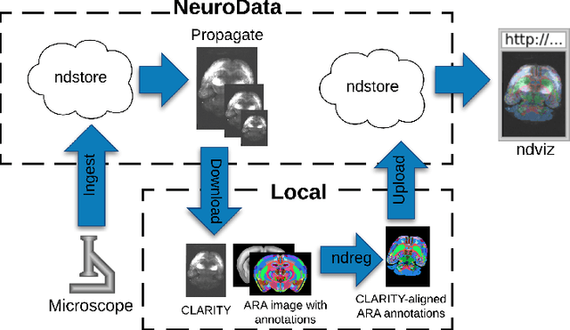

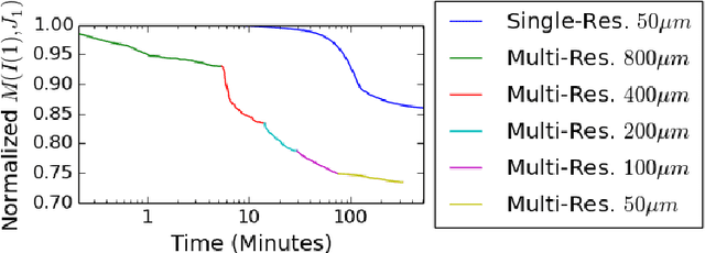
Abstract:CLARITY is a method for converting biological tissues into translucent and porous hydrogel-tissue hybrids. This facilitates interrogation with light sheet microscopy and penetration of molecular probes while avoiding physical slicing. In this work, we develop a pipeline for registering CLARIfied mouse brains to an annotated brain atlas. Due to the novelty of this microscopy technique it is impractical to use absolute intensity values to align these images to existing standard atlases. Thus we adopt a large deformation diffeomorphic approach for registering images via mutual information matching. Furthermore we show how a cascaded multi-resolution approach can improve registration quality while reducing algorithm run time. As acquired image volumes were over a terabyte in size, they were far too large for work on personal computers. Therefore the NeuroData computational infrastructure was deployed for multi-resolution storage and visualization of these images and aligned annotations on the web.
Deformably Registering and Annotating Whole CLARITY Brains to an Atlas via Masked LDDMM
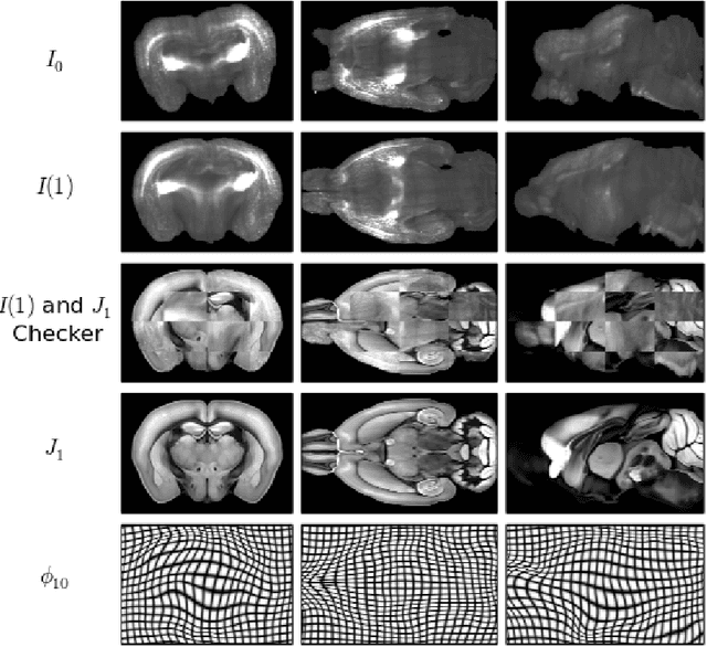
May 06, 2016Abstract:The CLARITY method renders brains optically transparent to enable high-resolution imaging in the structurally intact brain. Anatomically annotating CLARITY brains is necessary for discovering which regions contain signals of interest. Manually annotating whole-brain, terabyte CLARITY images is difficult, time-consuming, subjective, and error-prone. Automatically registering CLARITY images to a pre-annotated brain atlas offers a solution, but is difficult for several reasons. Removal of the brain from the skull and subsequent storage and processing cause variable non-rigid deformations, thus compounding inter-subject anatomical variability. Additionally, the signal in CLARITY images arises from various biochemical contrast agents which only sparsely label brain structures. This sparse labeling challenges the most commonly used registration algorithms that need to match image histogram statistics to the more densely labeled histological brain atlases. The standard method is a multiscale Mutual Information B-spline algorithm that dynamically generates an average template as an intermediate registration target. We determined that this method performs poorly when registering CLARITY brains to the Allen Institute's Mouse Reference Atlas (ARA), because the image histogram statistics are poorly matched. Therefore, we developed a method (Mask-LDDMM) for registering CLARITY images, that automatically find the brain boundary and learns the optimal deformation between the brain and atlas masks. Using Mask-LDDMM without an average template provided better results than the standard approach when registering CLARITY brains to the ARA. The LDDMM pipelines developed here provide a fast automated way to anatomically annotate CLARITY images. Our code is available as open source software at http://NeuroData.io.
 Add to Chrome
 Add to Firefox
 Add to Edge