Jia Dong
F3T: A soft tactile unit with 3D force and temperature mathematical decoupling ability for robots
Sep 05, 2024


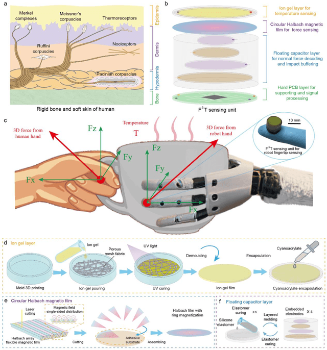
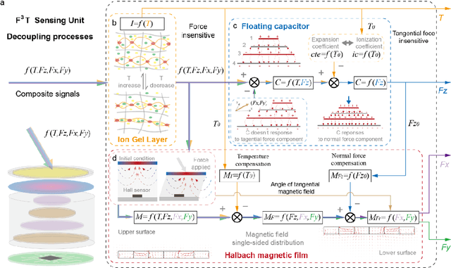
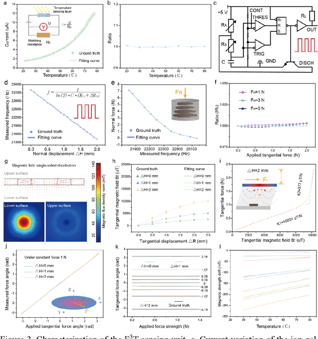
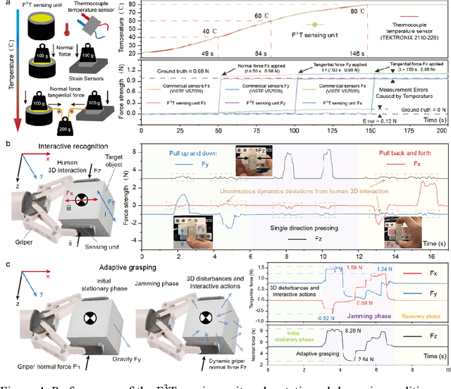
Abstract:The human skin exhibits remarkable capability to perceive contact forces and environmental temperatures, providing intricate information essential for nuanced manipulation. Despite recent advancements in soft tactile sensors, a significant challenge remains in accurately decoupling signals - specifically, separating force from directional orientation and temperature - resulting in fail to meet the advanced application requirements of robots. This research proposes a multi-layered soft sensor unit (F3T) designed to achieve isolated measurements and mathematical decoupling of normal pressure, omnidirectional tangential forces, and temperature. We developed a circular coaxial magnetic film featuring a floating-mountain multi-layer capacitor, facilitating the physical decoupling of normal and tangential forces in all directions. Additionally, we incorporated an ion gel-based temperature sensing film atop the tactile sensor. This sensor is resilient to external pressure and deformation, enabling it to measure temperature and, crucially, eliminate capacitor errors induced by environmental temperature changes. This innovative design allows for the decoupled measurement of multiple signals, paving the way for advancements in higher-level robot motion control, autonomous decision-making, and task planning.
From Pixel to Slide image: Polarization Modality-based Pathological Diagnosis Using Representation Learning
Jan 03, 2024


Abstract:Thyroid cancer is the most common endocrine malignancy, and accurately distinguishing between benign and malignant thyroid tumors is crucial for developing effective treatment plans in clinical practice. Pathologically, thyroid tumors pose diagnostic challenges due to improper specimen sampling. In this study, we have designed a three-stage model using representation learning to integrate pixel-level and slice-level annotations for distinguishing thyroid tumors. This structure includes a pathology structure recognition method to predict structures related to thyroid tumors, an encoder-decoder network to extract pixel-level annotation information by learning the feature representations of image blocks, and an attention-based learning mechanism for the final classification task. This mechanism learns the importance of different image blocks in a pathological region, globally considering the information from each block. In the third stage, all information from the image blocks in a region is aggregated using attention mechanisms, followed by classification to determine the category of the region. Experimental results demonstrate that our proposed method can predict microscopic structures more accurately. After color-coding, the method achieves results on unstained pathology slides that approximate the quality of Hematoxylin and eosin staining, reducing the need for stained pathology slides. Furthermore, by leveraging the concept of indirect measurement and extracting polarized features from structures correlated with lesions, the proposed method can also classify samples where membrane structures cannot be obtained through sampling, providing a potential objective and highly accurate indirect diagnostic technique for thyroid tumors.
A Polarization and Radiomics Feature Fusion Network for the Classification of Hepatocellular Carcinoma and Intrahepatic Cholangiocarcinoma
Dec 27, 2023Abstract:Classifying hepatocellular carcinoma (HCC) and intrahepatic cholangiocarcinoma (ICC) is a critical step in treatment selection and prognosis evaluation for patients with liver diseases. Traditional histopathological diagnosis poses challenges in this context. In this study, we introduce a novel polarization and radiomics feature fusion network, which combines polarization features obtained from Mueller matrix images of liver pathological samples with radiomics features derived from corresponding pathological images to classify HCC and ICC. Our fusion network integrates a two-tier fusion approach, comprising early feature-level fusion and late classification-level fusion. By harnessing the strengths of polarization imaging techniques and image feature-based machine learning, our proposed fusion network significantly enhances classification accuracy. Notably, even at reduced imaging resolutions, the fusion network maintains robust performance due to the additional information provided by polarization features, which may not align with human visual perception. Our experimental results underscore the potential of this fusion network as a powerful tool for computer-aided diagnosis of HCC and ICC, showcasing the benefits and prospects of integrating polarization imaging techniques into the current image-intensive digital pathological diagnosis. We aim to contribute this innovative approach to top-tier journals, offering fresh insights and valuable tools in the fields of medical imaging and cancer diagnosis. By introducing polarization imaging into liver cancer classification, we demonstrate its interdisciplinary potential in addressing challenges in medical image analysis, promising advancements in medical imaging and cancer diagnosis.
 Add to Chrome
 Add to Firefox
 Add to Edge