Hyuk-Jae Chang
A Unified Approach for Comprehensive Analysis of Various Spectral and Tissue Doppler Echocardiography
Nov 14, 2023
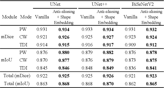
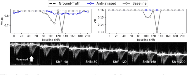
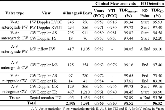
Abstract:Doppler echocardiography offers critical insights into cardiac function and phases by quantifying blood flow velocities and evaluating myocardial motion. However, previous methods for automating Doppler analysis, ranging from initial signal processing techniques to advanced deep learning approaches, have been constrained by their reliance on electrocardiogram (ECG) data and their inability to process Doppler views collectively. We introduce a novel unified framework using a convolutional neural network for comprehensive analysis of spectral and tissue Doppler echocardiography images that combines automatic measurements and end-diastole (ED) detection into a singular method. The network automatically recognizes key features across various Doppler views, with novel Doppler shape embedding and anti-aliasing modules enhancing interpretation and ensuring consistent analysis. Empirical results indicate a consistent outperformance in performance metrics, including dice similarity coefficients (DSC) and intersection over union (IoU). The proposed framework demonstrates strong agreement with clinicians in Doppler automatic measurements and competitive performance in ED detection.
Self supervised convolutional kernel based handcrafted feature harmonization: Enhanced left ventricle hypertension disease phenotyping on echocardiography
Oct 13, 2023Abstract:Radiomics, a medical imaging technique, extracts quantitative handcrafted features from images to predict diseases. Harmonization in those features ensures consistent feature extraction across various imaging devices and protocols. Methods for harmonization include standardized imaging protocols, statistical adjustments, and evaluating feature robustness. Myocardial diseases such as Left Ventricular Hypertrophy (LVH) and Hypertensive Heart Disease (HHD) are diagnosed via echocardiography, but variable imaging settings pose challenges. Harmonization techniques are crucial for applying handcrafted features in disease diagnosis in such scenario. Self-supervised learning (SSL) enhances data understanding within limited datasets and adapts to diverse data settings. ConvNeXt-V2 integrates convolutional layers into SSL, displaying superior performance in various tasks. This study focuses on convolutional filters within SSL, using them as preprocessing to convert images into feature maps for handcrafted feature harmonization. Our proposed method excelled in harmonization evaluation and exhibited superior LVH classification performance compared to existing methods.
Echocardiographic View Classification with Integrated Out-of-Distribution Detection for Enhanced Automatic Echocardiographic Analysis
Aug 31, 2023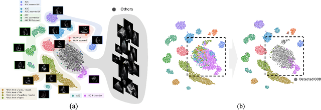


Abstract:In the rapidly evolving field of automatic echocardiographic analysis and interpretation, automatic view classification is a critical yet challenging task, owing to the inherent complexity and variability of echocardiographic data. This study presents ECHOcardiography VIew Classification with Out-of-Distribution dEtection (ECHO-VICODE), a novel deep learning-based framework that effectively addresses this challenge by training to classify 31 classes, surpassing previous studies and demonstrating its capacity to handle a wide range of echocardiographic views. Furthermore, ECHO-VICODE incorporates an integrated out-of-distribution (OOD) detection function, leveraging the relative Mahalanobis distance to effectively identify 'near-OOD' instances commonly encountered in echocardiographic data. Through extensive experimentation, we demonstrated the outstanding performance of ECHO-VICODE in terms of view classification and OOD detection, significantly reducing the potential for errors in echocardiographic analyses. This pioneering study significantly advances the domain of automated echocardiography analysis and exhibits promising prospects for substantial applications in extensive clinical research and practice.
Calcium Removal From Cardiac CT Images Using Deep Convolutional Neural Network
Feb 20, 2018
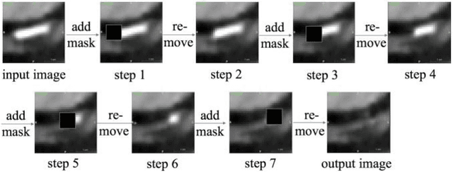
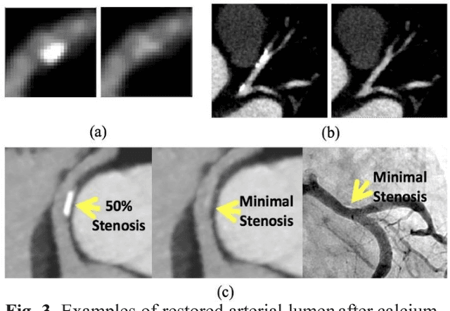
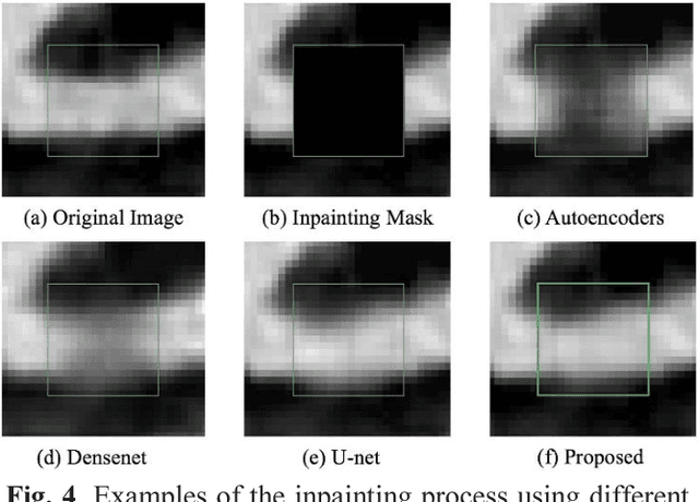
Abstract:Coronary calcium causes beam hardening and blooming artifacts on cardiac computed tomography angiography (CTA) images, which lead to overestimation of lumen stenosis and reduction of diagnostic specificity. To properly remove coronary calcification and restore arterial lumen precisely, we propose a machine learning-based method with a multi-step inpainting process. We developed a new network configuration, Dense-Unet, to achieve optimal performance with low computational cost. Results after the calcium removal process were validated by comparing with gold-standard X-ray angiography. Our results demonstrated that removing coronary calcification from images with the proposed approach was feasible, and may potentially improve the diagnostic accuracy of CTA.
 Add to Chrome
 Add to Firefox
 Add to Edge