Hongkun Sun
PSFHS Challenge Report: Pubic Symphysis and Fetal Head Segmentation from Intrapartum Ultrasound Images
Sep 17, 2024


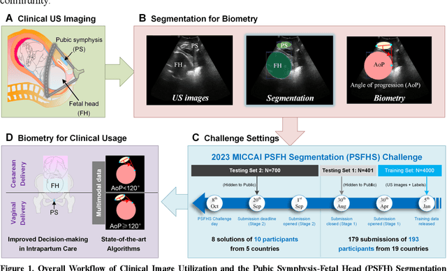
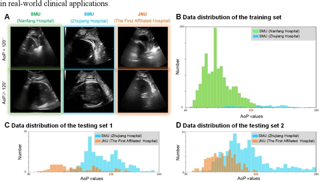
Abstract:Segmentation of the fetal and maternal structures, particularly intrapartum ultrasound imaging as advocated by the International Society of Ultrasound in Obstetrics and Gynecology (ISUOG) for monitoring labor progression, is a crucial first step for quantitative diagnosis and clinical decision-making. This requires specialized analysis by obstetrics professionals, in a task that i) is highly time- and cost-consuming and ii) often yields inconsistent results. The utility of automatic segmentation algorithms for biometry has been proven, though existing results remain suboptimal. To push forward advancements in this area, the Grand Challenge on Pubic Symphysis-Fetal Head Segmentation (PSFHS) was held alongside the 26th International Conference on Medical Image Computing and Computer Assisted Intervention (MICCAI 2023). This challenge aimed to enhance the development of automatic segmentation algorithms at an international scale, providing the largest dataset to date with 5,101 intrapartum ultrasound images collected from two ultrasound machines across three hospitals from two institutions. The scientific community's enthusiastic participation led to the selection of the top 8 out of 179 entries from 193 registrants in the initial phase to proceed to the competition's second stage. These algorithms have elevated the state-of-the-art in automatic PSFHS from intrapartum ultrasound images. A thorough analysis of the results pinpointed ongoing challenges in the field and outlined recommendations for future work. The top solutions and the complete dataset remain publicly available, fostering further advancements in automatic segmentation and biometry for intrapartum ultrasound imaging.
ParaTransCNN: Parallelized TransCNN Encoder for Medical Image Segmentation
Jan 27, 2024Abstract:The convolutional neural network-based methods have become more and more popular for medical image segmentation due to their outstanding performance. However, they struggle with capturing long-range dependencies, which are essential for accurately modeling global contextual correlations. Thanks to the ability to model long-range dependencies by expanding the receptive field, the transformer-based methods have gained prominence. Inspired by this, we propose an advanced 2D feature extraction method by combining the convolutional neural network and Transformer architectures. More specifically, we introduce a parallelized encoder structure, where one branch uses ResNet to extract local information from images, while the other branch uses Transformer to extract global information. Furthermore, we integrate pyramid structures into the Transformer to extract global information at varying resolutions, especially in intensive prediction tasks. To efficiently utilize the different information in the parallelized encoder at the decoder stage, we use a channel attention module to merge the features of the encoder and propagate them through skip connections and bottlenecks. Intensive numerical experiments are performed on both aortic vessel tree, cardiac, and multi-organ datasets. By comparing with state-of-the-art medical image segmentation methods, our method is shown with better segmentation accuracy, especially on small organs. The code is publicly available on https://github.com/HongkunSun/ParaTransCNN.
 Add to Chrome
 Add to Firefox
 Add to Edge