H. D. Cheng
Fuzzy Semantic Segmentation of Breast Ultrasound Image with Breast Anatomy Constraints
Oct 23, 2019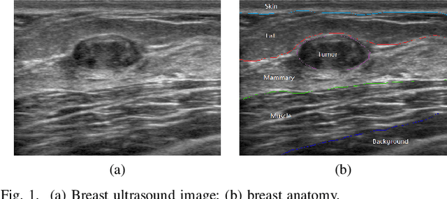
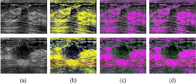

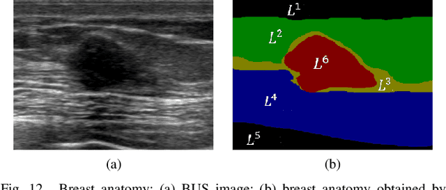
Abstract:Breast cancer is one of the most serious disease affecting women's health. Due to low cost, portable, no radiation, and high efficiency, breast ultrasound (BUS) imaging is the most popular approach for diagnosing early breast cancer. However, ultrasound images are low resolution and poor quality. Thus, developing accurate detection system is a challenging task. In this paper, we propose a fully automatic segmentation algorithm consisting of two parts: fuzzy fully convolutional network and accurately fine-tuning post-processing based on breast anatomy constraints. In the first part, the image is preprocessed by contrast enhancement, and wavelet features are employed for image augmentation. A fuzzy membership function transforms the augmented BUS images into fuzzy domain. The features from convolutional layers are processed using fuzzy logic as well. The conditional random fields (CRFs) post-process the segmentation result. The location relation among the breast anatomy layers is utilized to improve the performance. The proposed method is applied to the dataset with 325 BUS images, and achieves state-of-art performance compared with that of existing methods with true positive rate 90.33%, false positive rate 9.00%, and intersection over union (IoU) 81.29% on tumor category, and overall intersection over union (mIoU) 80.47% over five categories: fat layer, mammary layer, muscle layer, background, and tumor.
Breast Anatomy Enriched Tumor Saliency Estimation
Oct 23, 2019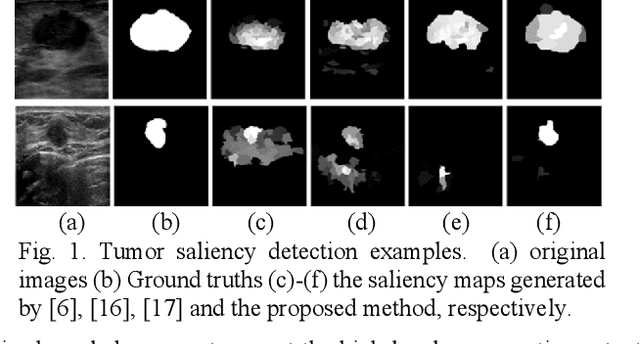
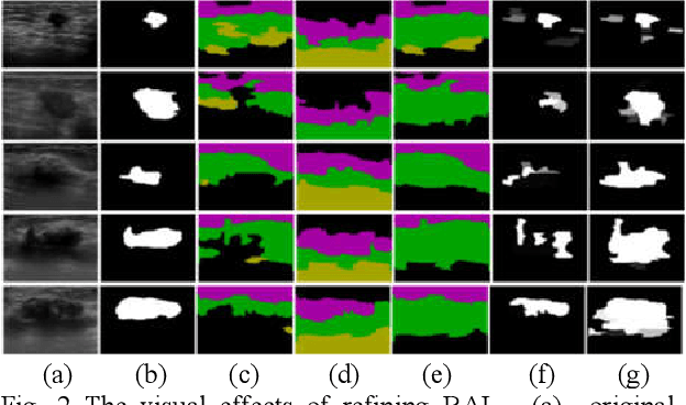
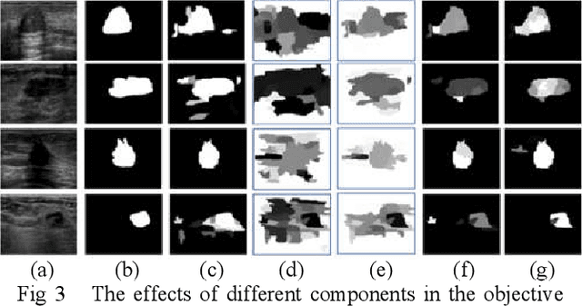
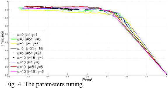
Abstract:Breast cancer investigation is of great significance, and developing tumor detection methodologies is a critical need. However, it is a challenging task for breast ultrasound due to the complicated breast structure and poor quality of the images. In this paper, we propose a novel tumor saliency estimation model guided by enriched breast anatomy knowledge to localize the tumor. Firstly, the breast anatomy layers are generated by a deep neural network. Then we refine the layers by integrating a non-semantic breast anatomy model to solve the problems of incomplete mammary layers. Meanwhile, a new background map generation method weighted by the semantic probability and spatial distance is proposed to improve the performance. The experiment demonstrates that the proposed method with the new background map outperforms four state-of-the-art TSE models with increasing 10% of F_meansure on the BUS public dataset.
Tumor Saliency Estimation for Breast Ultrasound Images via Breast Anatomy Modeling
Jun 18, 2019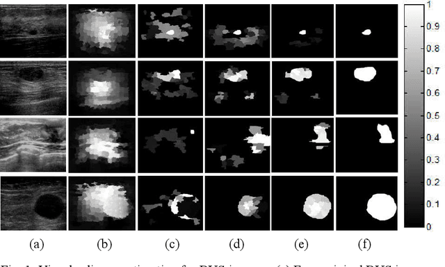
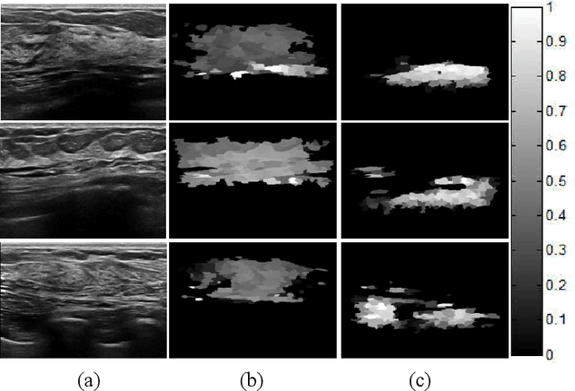
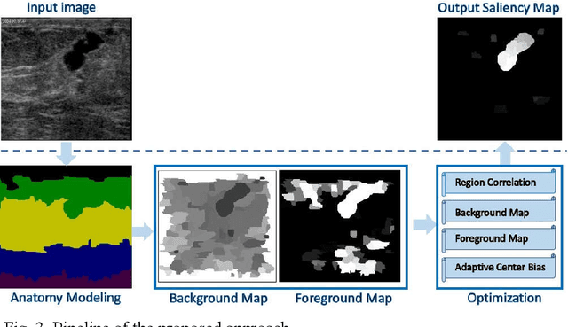
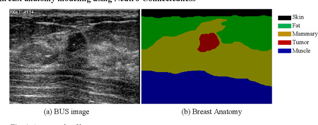
Abstract:Tumor saliency estimation aims to localize tumors by modeling the visual stimuli in medical images. However, it is a challenging task for breast ultrasound due to the complicated anatomic structure of the breast and poor image quality; and existing saliency estimation approaches only model generic visual stimuli, e.g., local and global contrast, location, and feature correlation, and achieve poor performance for tumor saliency estimation. In this paper, we propose a novel optimization model to estimate tumor saliency by utilizing breast anatomy. First, we model breast anatomy and decompose breast ultrasound image into layers using Neutro-Connectedness; then utilize the layers to generate the foreground and background maps; and finally propose a novel objective function to estimate the tumor saliency by integrating the foreground map, background map, adaptive center bias, and region-based correlation cues. The extensive experiments demonstrate that the proposed approach obtains more accurate foreground and background maps with the assistance of the breast anatomy; especially, for the images having large or small tumors; meanwhile, the new objective function can handle the images without tumors. The newly proposed method achieves state-of-the-art performance when compared to eight tumor saliency estimation approaches using two breast ultrasound datasets.
Abstaining Classification When Error Costs are Unequal and Unknown
Jul 25, 2018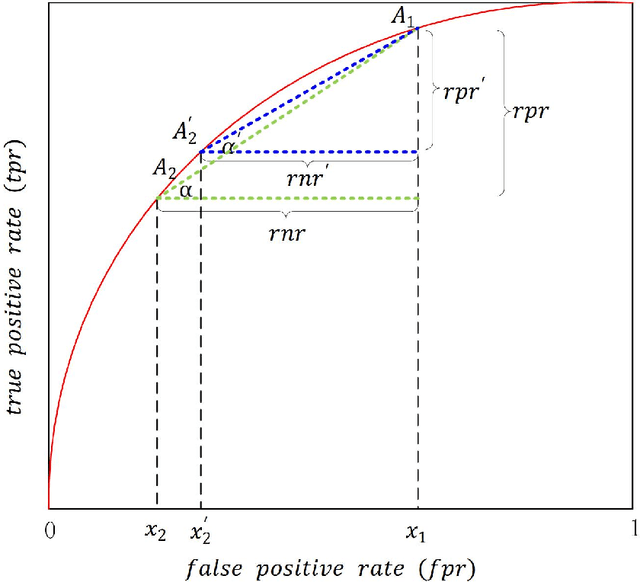

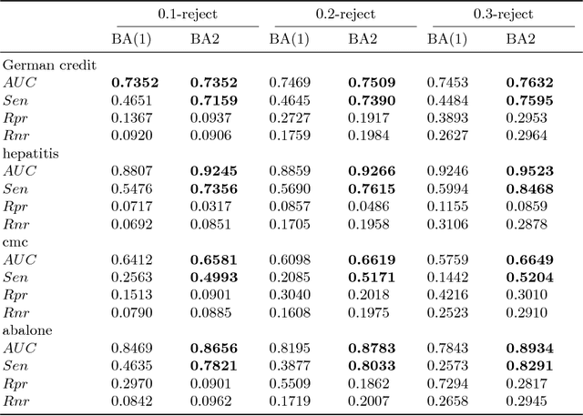
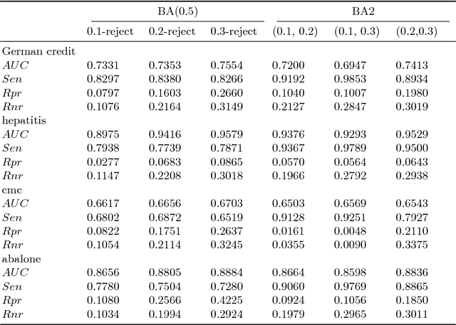
Abstract:Abstaining classificaiton aims to reject to classify the easily misclassified examples, so it is an effective approach to increase the clasificaiton reliability and reduce the misclassification risk in the cost-sensitive applications. In such applications, different types of errors (false positive or false negative) usaully have unequal costs. And the error costs, which depend on specific applications, are usually unknown. However, current abstaining classification methods either do not distinguish the error types, or they need the cost information of misclassification and rejection, which are realized in the framework of cost-sensitive learning. In this paper, we propose a bounded-abstention method with two constraints of reject rates (BA2), which performs abstaining classification when error costs are unequal and unknown. BA2 aims to obtain the optimal area under the ROC curve (AUC) by constraining the reject rates of the positive and negative classes respectively. Specifically, we construct the receiver operating characteristic (ROC) curve, and stepwise search the optimal reject thresholds from both ends of the curve, untill the two constraints are satisfied. Experimental results show that BA2 obtains higher AUC and lower total cost than the state-of-the-art abstaining classification methods. Meanwhile, BA2 achieves controllable reject rates of the positive and negative classes.
A Hybrid Framework for Tumor Saliency Estimation
Jun 27, 2018


Abstract:Automatic tumor segmentation of breast ultrasound (BUS) image is quite challenging due to the complicated anatomic structure of breast and poor image quality. Most tumor segmentation approaches achieve good performance on BUS images collected in controlled settings; however, the performance degrades greatly with BUS images from different sources. Tumor saliency estimation (TSE) has attracted increasing attention to solving the problem by modeling radiologists' attention mechanism. In this paper, we propose a novel hybrid framework for TSE, which integrates both high-level domain-knowledge and robust low-level saliency assumptions and can overcome drawbacks caused by direct mapping in traditional TSE approaches. The new framework integrated the Neutro-Connectedness (NC) map, the adaptive-center, the correlation and the layer structure-based weighted map. The experimental results demonstrate that the proposed approach outperforms state-of-the-art TSE methods.
Computer-Aided Knee Joint Magnetic Resonance Image Segmentation - A Survey
Feb 13, 2018


Abstract:Osteoarthritis (OA) is one of the major health issues among the elderly population. MRI is the most popular technology to observe and evaluate the progress of OA course. However, the extreme labor cost of MRI analysis makes the process inefficient and expensive. Also, due to human error and subjective nature, the inter- and intra-observer variability is rather high. Computer-aided knee MRI segmentation is currently an active research field because it can alleviate doctors and radiologists from the time consuming and tedious job, and improve the diagnosis performance which has immense potential for both clinic and scientific research. In the past decades, researchers have investigated automatic/semi-automatic knee MRI segmentation methods extensively. However, to the best of our knowledge, there is no comprehensive survey paper in this field yet. In this survey paper, we classify the existing methods by their principles and discuss the current research status and point out the future research trend in-depth.
Automatic Breast Ultrasound Image Segmentation: A Survey
Jan 09, 2018
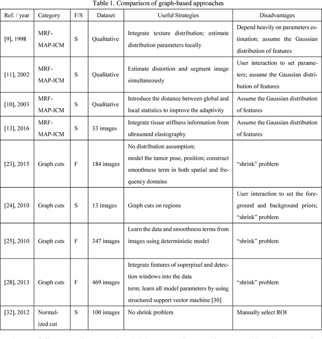


Abstract:Breast cancer is one of the leading causes of cancer death among women worldwide. In clinical routine, automatic breast ultrasound (BUS) image segmentation is very challenging and essential for cancer diagnosis and treatment planning. Many BUS segmentation approaches have been studied in the last two decades, and have been proved to be effective on private datasets. Currently, the advancement of BUS image segmentation seems to meet its bottleneck. The improvement of the performance is increasingly challenging, and only few new approaches were published in the last several years. It is the time to look at the field by reviewing previous approaches comprehensively and to investigate the future directions. In this paper, we study the basic ideas, theories, pros and cons of the approaches, group them into categories, and extensively review each category in depth by discussing the principles, application issues, and advantages/disadvantages.
A Benchmark for Breast Ultrasound Image Segmentation (BUSIS)
Jan 09, 2018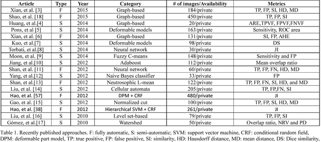
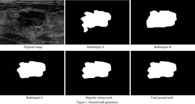

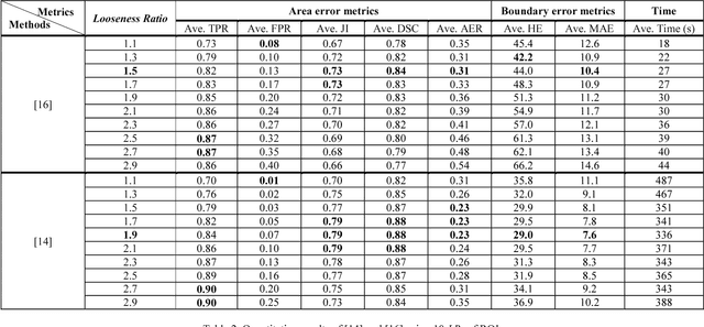
Abstract:Breast ultrasound (BUS) image segmentation is challenging and critical for BUS Computer-Aided Diagnosis (CAD) systems. Many BUS segmentation approaches have been proposed in the last two decades, but the performances of most approaches have been assessed using relatively small private datasets with differ-ent quantitative metrics, which result in discrepancy in performance comparison. Therefore, there is a pressing need for building a benchmark to compare existing methods using a public dataset objectively, and to determine the performance of the best breast tumor segmentation algorithm available today and to investigate what segmentation strategies are valuable in clinical practice and theoretical study. In this work, we will publish a B-mode BUS image segmentation benchmark (BUSIS) with 562 images and compare the performance of five state-of-the-art BUS segmentation methods quantitatively.
Neutro-Connectedness Cut
Aug 06, 2016


Abstract:Interactive image segmentation is a challenging task and receives increasing attention recently; however, two major drawbacks exist in interactive segmentation approaches. First, the segmentation performance of ROI-based methods is sensitive to the initial ROI: different ROIs may produce results with great difference. Second, most seed-based methods need intense interactions, and are not applicable in many cases. In this work, we generalize the Neutro-Connectedness (NC) to be independent of top-down priors of objects and to model image topology with indeterminacy measurement on image regions, propose a novel method for determining object and background regions, which is applied to exclude isolated background regions and enforce label consistency, and put forward a hybrid interactive segmentation method, Neutro-Connectedness Cut (NC-Cut), which can overcome the above two problems by utilizing both pixel-wise appearance information and region-based NC properties. We evaluate the proposed NC-Cut by employing two image datasets (265 images), and demonstrate that the proposed approach outperforms state-of-the-art interactive image segmentation methods (Grabcut, MILCut, One-Cut, MGC_max^sum and pPBC).
An algorithm for Left Atrial Thrombi detection using Transesophageal Echocardiography
Aug 24, 2015


Abstract:Transesophageal echocardiography (TEE) is widely used to detect left atrium (LA)/left atrial appendage (LAA) thrombi. In this paper, the local binary pattern variance (LBPV) features are extracted from region of interest (ROI). And the dynamic features are formed by using the information of its neighbor frames in the sequence. The sequence is viewed as a bag, and the images in the sequence are considered as the instances. Multiple-instance learning (MIL) method is employed to solve the LAA thrombi detection. The experimental results show that the proposed method can achieve better performance than that by using other methods.
 Add to Chrome
 Add to Firefox
 Add to Edge