Gouenou Coatrieux
FedCLEAN: byzantine defense by CLustering Errors of Activation maps in Non-IID federated learning environments
Jan 21, 2025Abstract:Federated Learning (FL) enables clients to collaboratively train a global model using their local datasets while reinforcing data privacy. However, FL is susceptible to poisoning attacks. Existing defense mechanisms assume that clients' data are independent and identically distributed (IID), making them ineffective in real-world applications where data are non-IID. This paper presents FedCLEAN, the first defense capable of filtering attackers' model updates in a non-IID FL environment. The originality of FedCLEAN is twofold. First, it relies on a client confidence score derived from the reconstruction errors of each client's model activation maps for a given trigger set, with reconstruction errors obtained by means of a Conditional Variational Autoencoder trained according to a novel server-side strategy. Second, we propose an ad-hoc trust propagation algorithm based on client scores, which allows building a cluster of benign clients while flagging potential attackers. Experimental results on the datasets MNIST and FashionMNIST demonstrate the robustness of FedCLEAN against Byzantine attackers in non-IID scenarios and a close-to-zero benign client misclassification rate, even in the absence of an attack.
When Federated Learning meets Watermarking: A Comprehensive Overview of Techniques for Intellectual Property Protection
Aug 07, 2023


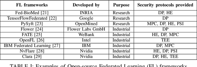
Abstract:Federated Learning (FL) is a technique that allows multiple participants to collaboratively train a Deep Neural Network (DNN) without the need of centralizing their data. Among other advantages, it comes with privacy-preserving properties making it attractive for application in sensitive contexts, such as health care or the military. Although the data are not explicitly exchanged, the training procedure requires sharing information about participants' models. This makes the individual models vulnerable to theft or unauthorized distribution by malicious actors. To address the issue of ownership rights protection in the context of Machine Learning (ML), DNN Watermarking methods have been developed during the last five years. Most existing works have focused on watermarking in a centralized manner, but only a few methods have been designed for FL and its unique constraints. In this paper, we provide an overview of recent advancements in Federated Learning watermarking, shedding light on the new challenges and opportunities that arise in this field.
White-box Membership Attack Against Machine Learning Based Retinopathy Classification
May 30, 2022

Abstract:The advances in machine learning (ML) have greatly improved AI-based diagnosis aid systems in medical imaging. However, being based on collecting medical data specific to individuals induces several security issues, especially in terms of privacy. Even though the owner of the images like a hospital put in place strict privacy protection provisions at the level of its information system, the model trained over his images still holds disclosure potential. The trained model may be accessible to an attacker as: 1) White-box: accessing to the model architecture and parameters; 2) Black box: where he can only query the model with his own inputs through an appropriate interface. Existing attack methods include: feature estimation attacks (FEA), membership inference attack (MIA), model memorization attack (MMA) and identification attacks (IA). In this work we focus on MIA against a model that has been trained to detect diabetic retinopathy from retinal images. Diabetic retinopathy is a condition that can cause vision loss and blindness in the people who have diabetes. MIA is the process of determining whether a data sample comes from the training data set of a trained ML model or not. From a privacy perspective in our use case where a diabetic retinopathy classification model is given to partners that have at their disposal images along with patients' identifiers, inferring the membership status of a data sample can help to state if a patient has contributed or not to the training of the model.
Two-stage breast mass detection and segmentation system towards automated high-resolution full mammogram analysis
Feb 27, 2020


Abstract:Mammography is the primary imaging modality used for early detection and diagnosis of breast cancer. Mammography analysis mainly refers to the extraction of regions of interest around tumors, followed by a segmentation step, which is essential to further classification of benign or malignant tumors. Breast masses are the most important findings among breast abnormalities. However, manual delineation of masses from native mammogram is a time consuming and error-prone task. An integrated computer-aided diagnosis system to assist radiologists in automatically detecting and segmenting breast masses is therefore in urgent need. We propose a fully-automated approach that guides accurate mass segmentation from full mammograms at high resolution through a detection stage. First, mass detection is performed by an efficient deep learning approach, You-Only-Look-Once, extended by integrating multi-scale predictions to improve automatic candidate selection. Second, a convolutional encoder-decoder network using nested and dense skip connections is employed to fine-delineate candidate masses. Unlike most previous studies based on segmentation from regions, our framework handles mass segmentation from native full mammograms without user intervention. Trained on INbreast and DDSM-CBIS public datasets, the pipeline achieves an overall average Dice of 80.44% on high-resolution INbreast test images, outperforming state-of-the-art methods. Our system shows promising accuracy as an automatic full-image mass segmentation system. The comprehensive evaluation provided for both detection and segmentation stages reveals strong robustness to the diversity of size, shape and appearance of breast masses, towards better computer-aided diagnosis.
Real-time analysis of cataract surgery videos using statistical models
Oct 18, 2016


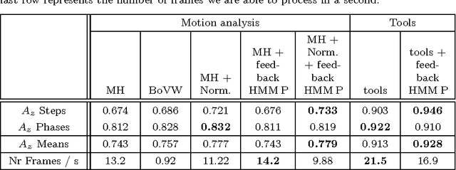
Abstract:The automatic analysis of the surgical process, from videos recorded during surgeries, could be very useful to surgeons, both for training and for acquiring new techniques. The training process could be optimized by automatically providing some targeted recommendations or warnings, similar to the expert surgeon's guidance. In this paper, we propose to reuse videos recorded and stored during cataract surgeries to perform the analysis. The proposed system allows to automatically recognize, in real time, what the surgeon is doing: what surgical phase or, more precisely, what surgical step he or she is performing. This recognition relies on the inference of a multilevel statistical model which uses 1) the conditional relations between levels of description (steps and phases) and 2) the temporal relations among steps and among phases. The model accepts two types of inputs: 1) the presence of surgical tools, manually provided by the surgeons, or 2) motion in videos, automatically analyzed through the Content Based Video retrieval (CBVR) paradigm. Different data-driven statistical models are evaluated in this paper. For this project, a dataset of 30 cataract surgery videos was collected at Brest University hospital. The system was evaluated in terms of area under the ROC curve. Promising results were obtained using either the presence of surgical tools ($A_z$ = 0.983) or motion analysis ($A_z$ = 0.759). The generality of the method allows to adapt it to any kinds of surgeries. The proposed solution could be used in a computer assisted surgery tool to support surgeons during the surgery.
 Add to Chrome
 Add to Firefox
 Add to Edge