Katia Charrière
Deep image mining for diabetic retinopathy screening
Apr 28, 2017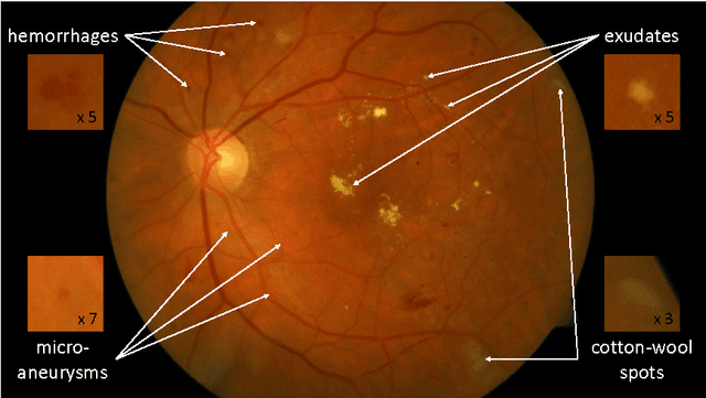
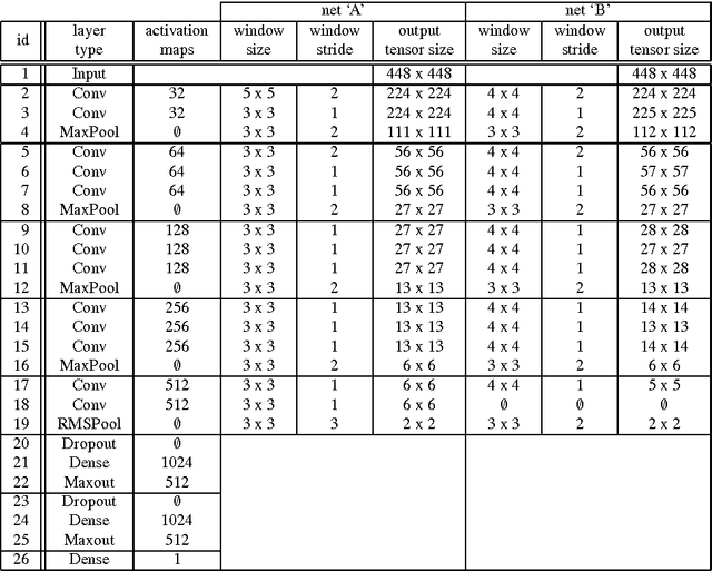
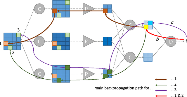
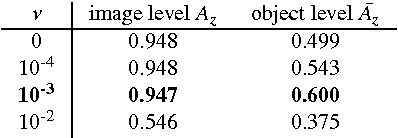
Abstract:Deep learning is quickly becoming the leading methodology for medical image analysis. Given a large medical archive, where each image is associated with a diagnosis, efficient pathology detectors or classifiers can be trained with virtually no expert knowledge about the target pathologies. However, deep learning algorithms, including the popular ConvNets, are black boxes: little is known about the local patterns analyzed by ConvNets to make a decision at the image level. A solution is proposed in this paper to create heatmaps showing which pixels in images play a role in the image-level predictions. In other words, a ConvNet trained for image-level classification can be used to detect lesions as well. A generalization of the backpropagation method is proposed in order to train ConvNets that produce high-quality heatmaps. The proposed solution is applied to diabetic retinopathy (DR) screening in a dataset of almost 90,000 fundus photographs from the 2015 Kaggle Diabetic Retinopathy competition and a private dataset of almost 110,000 photographs (e-ophtha). For the task of detecting referable DR, very good detection performance was achieved: $A_z = 0.954$ in Kaggle's dataset and $A_z = 0.949$ in e-ophtha. Performance was also evaluated at the image level and at the lesion level in the DiaretDB1 dataset, where four types of lesions are manually segmented: microaneurysms, hemorrhages, exudates and cotton-wool spots. The proposed detector outperforms recent algorithms trained to detect those lesions specifically, as well as competing heatmap generation algorithms for ConvNets. This detector is part of the Messidor system for mobile eye pathology screening. Because it does not rely on expert knowledge or manual segmentation for detecting relevant patterns, the proposed solution is a promising image mining tool, which has the potential to discover new biomarkers in images.
* Accepted for publication in Medical Image Analysis
Real-time analysis of cataract surgery videos using statistical models
Oct 18, 2016
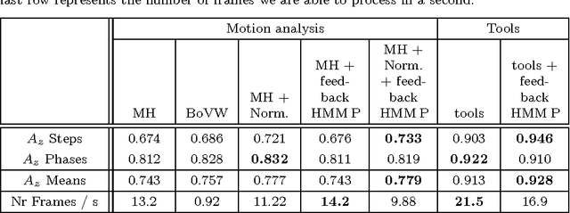


Abstract:The automatic analysis of the surgical process, from videos recorded during surgeries, could be very useful to surgeons, both for training and for acquiring new techniques. The training process could be optimized by automatically providing some targeted recommendations or warnings, similar to the expert surgeon's guidance. In this paper, we propose to reuse videos recorded and stored during cataract surgeries to perform the analysis. The proposed system allows to automatically recognize, in real time, what the surgeon is doing: what surgical phase or, more precisely, what surgical step he or she is performing. This recognition relies on the inference of a multilevel statistical model which uses 1) the conditional relations between levels of description (steps and phases) and 2) the temporal relations among steps and among phases. The model accepts two types of inputs: 1) the presence of surgical tools, manually provided by the surgeons, or 2) motion in videos, automatically analyzed through the Content Based Video retrieval (CBVR) paradigm. Different data-driven statistical models are evaluated in this paper. For this project, a dataset of 30 cataract surgery videos was collected at Brest University hospital. The system was evaluated in terms of area under the ROC curve. Promising results were obtained using either the presence of surgical tools ($A_z$ = 0.983) or motion analysis ($A_z$ = 0.759). The generality of the method allows to adapt it to any kinds of surgeries. The proposed solution could be used in a computer assisted surgery tool to support surgeons during the surgery.
 Add to Chrome
 Add to Firefox
 Add to Edge