Fedra Hajizadeh
Multi-Scale Convolutional Neural Network for Automated AMD Classification using Retinal OCT Images
Oct 06, 2021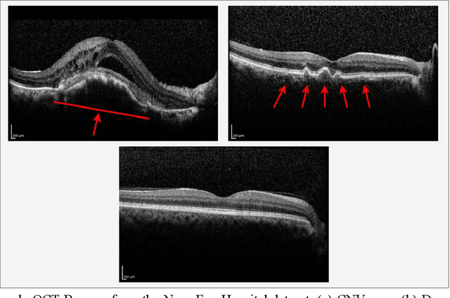

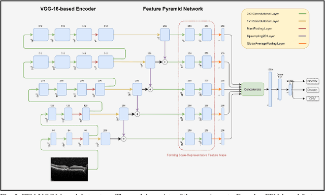
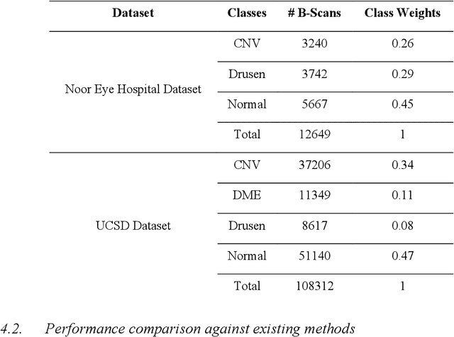
Abstract:Age-related macular degeneration (AMD) is the most common cause of blindness in developed countries, especially in people over 60 years of age. The workload of specialists and the healthcare system in this field has increased in recent years mainly dues to three reasons: 1) increased use of retinal optical coherence tomography (OCT) imaging technique, 2) prevalence of population aging worldwide, and 3) chronic nature of AMD. Recent developments in deep learning have provided a unique opportunity for the development of fully automated diagnosis frameworks. Considering the presence of AMD-related retinal pathologies in varying sizes in OCT images, our objective was to propose a multi-scale convolutional neural network (CNN) capable of distinguishing pathologies using receptive fields with various sizes. The multi-scale CNN was designed based on the feature pyramid network (FPN) structure and was used to diagnose normal and two common clinical characteristics of dry and wet AMD, namely drusen and choroidal neovascularization (CNV). The proposed method was evaluated on a national dataset gathered at Noor Eye Hospital (NEH), consisting of 12649 retinal OCT images from 441 patients, and a UCSD public dataset, consisting of 108312 OCT images. The results show that the multi-scale FPN-based structure was able to improve the base model's overall accuracy by 0.4% to 3.3% for different backbone models. In addition, gradual learning improved the performance in two phases from 87.2%+-2.5% to 93.4%+-1.4% by pre-training the base model on ImageNet weights in the first phase and fine-tuning the resulting model on a dataset of OCT images in the second phase. The promising quantitative and qualitative results of the proposed architecture prove the suitability of the proposed method to be used as a screening tool in healthcare centers assisting ophthalmologists in making better diagnostic decisions.
Thickness Mapping of Eleven Retinal Layers in Normal Eyes Using Spectral Domain Optical Coherence Tomography
Dec 11, 2013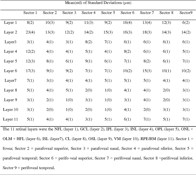
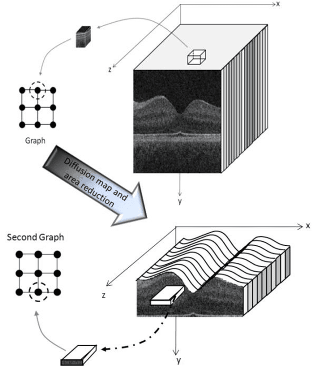
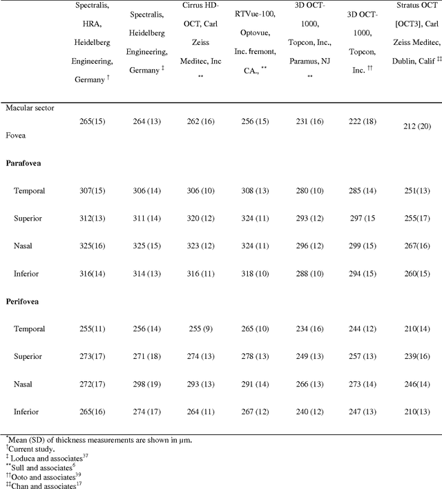
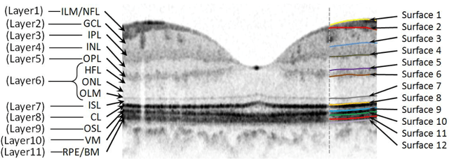
Abstract:Purpose. This study was conducted to determine the thickness map of eleven retinal layers in normal subjects by spectral domain optical coherence tomography (SD-OCT) and evaluate their association with sex and age. Methods. Mean regional retinal thickness of 11 retinal layers were obtained by automatic three-dimensional diffusion-map-based method in 112 normal eyes of 76 Iranian subjects. Results. The thickness map of central foveal area in layer 1, 3, and 4 displayed the minimum thickness (P<0.005 for all). Maximum thickness was observed in nasal to the fovea of layer 1 (P<0.001) and in a circular pattern in the parafoveal retinal area of layers 2, 3 and 4 and in central foveal area of layer 6 (P<0.001). Temporal and inferior quadrants of the total retinal thickness and most of other quadrants of layer 1 were significantly greater in the men than in the women. Surrounding eight sectors of total retinal thickness and a limited number of sectors in layer 1 and 4 significantly correlated with age. Conclusion. SD-OCT demonstrated the three-dimensional thickness distribution of retinal layers in normal eyes. Thickness of layers varied with sex and age and in different sectors. These variables should be considered while evaluating macular thickness.
 Add to Chrome
 Add to Firefox
 Add to Edge