David Hawkes
Evaluation of automated airway morphological quantification for assessing fibrosing lung disease
Nov 19, 2021


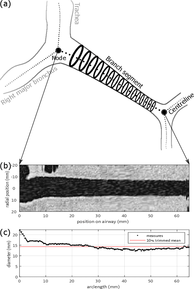
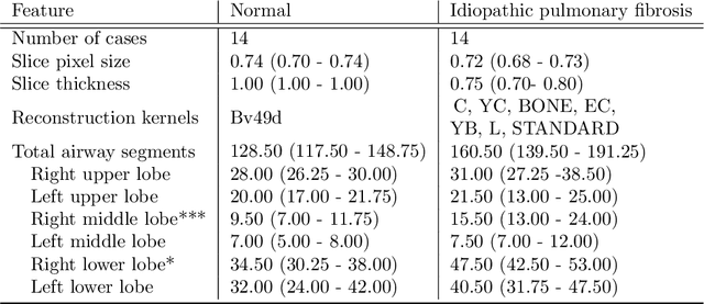
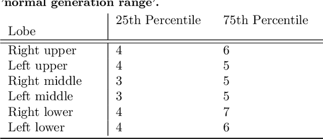
Abstract:Abnormal airway dilatation, termed traction bronchiectasis, is a typical feature of idiopathic pulmonary fibrosis (IPF). Volumetric computed tomography (CT) imaging captures the loss of normal airway tapering in IPF. We postulated that automated quantification of airway abnormalities could provide estimates of IPF disease extent and severity. We propose AirQuant, an automated computational pipeline that systematically parcellates the airway tree into its lobes and generational branches from a deep learning based airway segmentation, deriving airway structural measures from chest CT. Importantly, AirQuant prevents the occurrence of spurious airway branches by thick wave propagation and removes loops in the airway-tree by graph search, overcoming limitations of existing airway skeletonisation algorithms. Tapering between airway segments (intertapering) and airway tortuosity computed by AirQuant were compared between 14 healthy participants and 14 IPF patients. Airway intertapering was significantly reduced in IPF patients, and airway tortuosity was significantly increased when compared to healthy controls. Differences were most marked in the lower lobes, conforming to the typical distribution of IPF-related damage. AirQuant is an open-source pipeline that avoids limitations of existing airway quantification algorithms and has clinical interpretability. Automated airway measurements may have potential as novel imaging biomarkers of IPF severity and disease extent.
Pre-Quantized Deep Learning Models Codified in ONNX to Enable Hardware/Software Co-Design
Oct 04, 2021


Abstract:This paper presents a methodology to separate the quantization process from the hardware-specific model compilation stage via a pre-quantized deep learning model description in standard ONNX format. Separating the quantization process from the model compilation stage enables independent development. The methodology is expressive to convey hardware-specific operations and to embed key quantization parameters into a ONNX model which enables hardware/software co-design. Detailed examples are given for both MLP and CNN based networks, which can be extended to other networks in a straightforward fashion.
Modelling Airway Geometry as Stock Market Data using Bayesian Changepoint Detection
Jun 28, 2019


Abstract:Numerous lung diseases, such as idiopathic pulmonary fibrosis (IPF), exhibit dilation of the airways. Accurate measurement of dilatation enables assessment of the progression of disease. Unfortunately the combination of image noise and airway bifurcations causes high variability in the profiles of cross-sectional areas, rendering the identification of affected regions very difficult. Here we introduce a noise-robust method for automatically detecting the location of progressive airway dilatation given two profiles of the same airway acquired at different time points. We propose a probabilistic model of abrupt relative variations between profiles and perform inference via Reversible Jump Markov Chain Monte Carlo sampling. We demonstrate the efficacy of the proposed method on two datasets; (i) images of healthy airways with simulated dilatation; (ii) pairs of real images of IPF-affected airways acquired at 1 year intervals. Our model is able to detect the starting location of airway dilatation with an accuracy of 2.5mm on simulated data. The experiments on the IPF dataset display reasonable agreement with radiologists. We can compute a relative change in airway volume that may be useful for quantifying IPF disease progression.
Nonrigid reconstruction of 3D breast surfaces with a low-cost RGBD camera for surgical planning and aesthetic evaluation
Jan 16, 2019


Abstract:Accounting for 26% of all new cancer cases worldwide, breast cancer remains the most common form of cancer in women. Although early breast cancer has a favourable long-term prognosis, roughly a third of patients suffer from a suboptimal aesthetic outcome despite breast conserving cancer treatment. Clinical-quality 3D modelling of the breast surface therefore assumes an increasingly important role in advancing treatment planning, prediction and evaluation of breast cosmesis. Yet, existing 3D torso scanners are expensive and either infrastructure-heavy or subject to motion artefacts. In this paper we employ a single consumer-grade RGBD camera with an ICP-based registration approach to jointly align all points from a sequence of depth images non-rigidly. Subtle body deformation due to postural sway and respiration is successfully mitigated leading to a higher geometric accuracy through regularised locally affine transformations. We present results from 6 clinical cases where our method compares well with the gold standard and outperforms a previous approach. We show that our method produces better reconstructions qualitatively by visual assessment and quantitatively by consistently obtaining lower landmark error scores and yielding more accurate breast volume estimates.
A Log-Euclidean and Total Variation based Variational Framework for Computational Sonography
Feb 06, 2018Abstract:We propose a spatial compounding technique and variational framework to improve 3D ultrasound image quality by compositing multiple ultrasound volumes acquired from different probe orientations. In the composite volume, instead of intensity values, we estimate a tensor at every voxel. The resultant tensor image encapsulates the directional information of the underlying imaging data and can be used to generate ultrasound volumes from arbitrary, potentially unseen, probe positions. Extending the work of Hennersperger et al., we introduce a log-Euclidean framework to ensure that the tensors are positive-definite, eventually ensuring non-negative images. Additionally, we regularise the underpinning ill-posed variational problem while preserving edge information by relying on a total variation penalisation of the tensor field in the log domain. We present results on in vivo human data to show the efficacy of the approach.
A comparative study of breast surface reconstruction for aesthetic outcome assessment
Jun 20, 2017


Abstract:Breast cancer is the most prevalent cancer type in women, and while its survival rate is generally high the aesthetic outcome is an increasingly important factor when evaluating different treatment alternatives. 3D scanning and reconstruction techniques offer a flexible tool for building detailed and accurate 3D breast models that can be used both pre-operatively for surgical planning and post-operatively for aesthetic evaluation. This paper aims at comparing the accuracy of low-cost 3D scanning technologies with the significantly more expensive state-of-the-art 3D commercial scanners in the context of breast 3D reconstruction. We present results from 28 synthetic and clinical RGBD sequences, including 12 unique patients and an anthropomorphic phantom demonstrating the applicability of low-cost RGBD sensors to real clinical cases. Body deformation and homogeneous skin texture pose challenges to the studied reconstruction systems. Although these should be addressed appropriately if higher model quality is warranted, we observe that low-cost sensors are able to obtain valuable reconstructions comparable to the state-of-the-art within an error margin of 3 mm.
 Add to Chrome
 Add to Firefox
 Add to Edge