Daniel Stromer
Pattern Recognition Lab, FAU Erlangen-Nürnberg, Erlangen
Automatic Classification of Neuromuscular Diseases in Children Using Photoacoustic Imaging
Jan 27, 2022
Abstract:Neuromuscular diseases (NMDs) cause a significant burden for both healthcare systems and society. They can lead to severe progressive muscle weakness, muscle degeneration, contracture, deformity and progressive disability. The NMDs evaluated in this study often manifest in early childhood. As subtypes of disease, e.g. Duchenne Muscular Dystropy (DMD) and Spinal Muscular Atrophy (SMA), are difficult to differentiate at the beginning and worsen quickly, fast and reliable differential diagnosis is crucial. Photoacoustic and ultrasound imaging has shown great potential to visualize and quantify the extent of different diseases. The addition of automatic classification of such image data could further improve standard diagnostic procedures. We compare deep learning-based 2-class and 3-class classifiers based on VGG16 for differentiating healthy from diseased muscular tissue. This work shows promising results with high accuracies above 0.86 for the 3-class problem and can be used as a proof of concept for future approaches for earlier diagnosis and therapeutic monitoring of NMDs.
Initial Investigations Towards Non-invasive Monitoring of Chronic Wound Healing Using Deep Learning and Ultrasound Imaging
Jan 25, 2022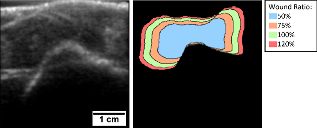

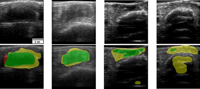
Abstract:Chronic wounds including diabetic and arterial/venous insufficiency injuries have become a major burden for healthcare systems worldwide. Demographic changes suggest that wound care will play an even bigger role in the coming decades. Predicting and monitoring response to therapy in wound care is currently largely based on visual inspection with little information on the underlying tissue. Thus, there is an urgent unmet need for innovative approaches that facilitate personalized diagnostics and treatments at the point-of-care. It has been recently shown that ultrasound imaging can monitor response to therapy in wound care, but this work required onerous manual image annotations. In this study, we present initial results of a deep learning-based automatic segmentation of cross-sectional wound size in ultrasound images and identify requirements and challenges for future research on this application. Evaluation of the segmentation results underscores the potential of the proposed deep learning approach to complement non-invasive imaging with Dice scores of 0.34 (U-Net, FCN) and 0.27 (ResNet-U-Net) but also highlights the need for improving robustness further. We conclude that deep learning-supported analysis of non-invasive ultrasound images is a promising area of research to automatically extract cross-sectional wound size and depth information with potential value in monitoring response to therapy.
Maximum a posteriori signal recovery for optical coherence tomography angiography image generation and denoising
Oct 29, 2020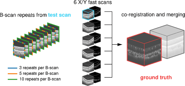
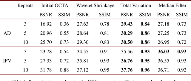
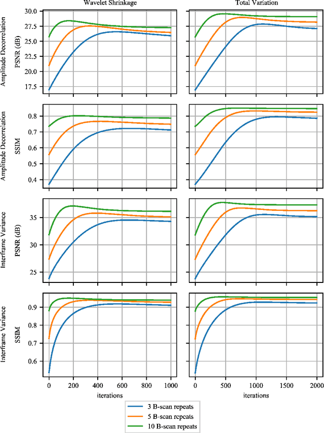
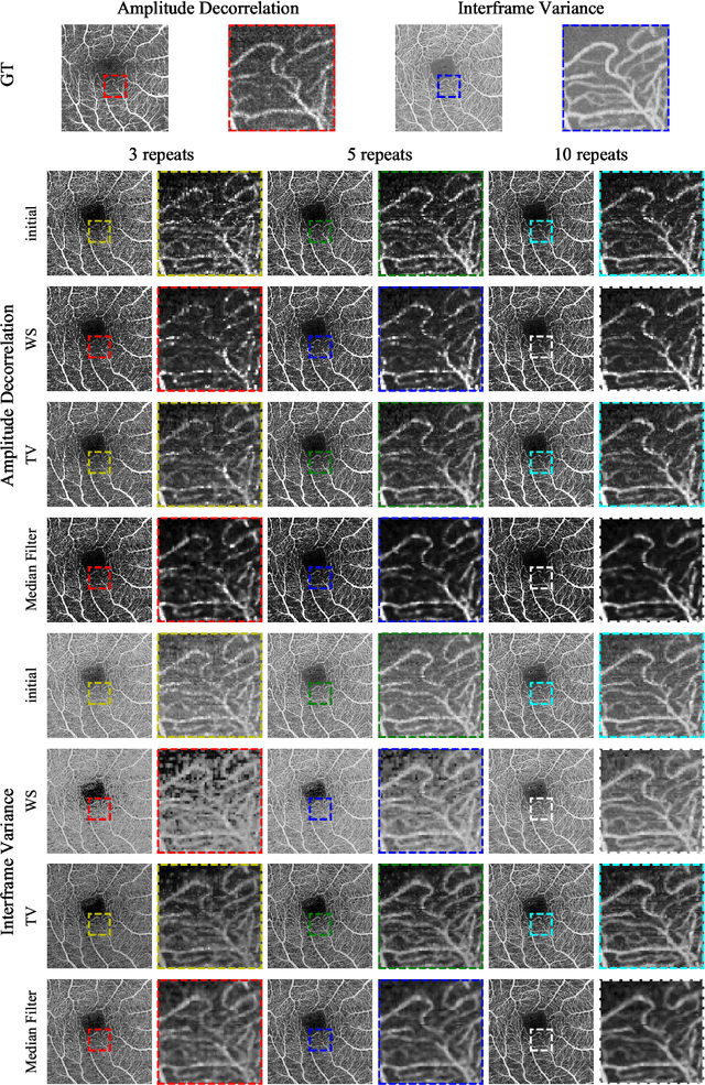
Abstract:Optical coherence tomography angiography (OCTA) is a novel and clinically promising imaging modality to image retinal and sub-retinal vasculature. Based on repeated optical coherence tomography (OCT) scans, intensity changes are observed over time and used to compute OCTA image data. OCTA data are prone to noise and artifacts caused by variations in flow speed and patient movement. We propose a novel iterative maximum a posteriori signal recovery algorithm in order to generate OCTA volumes with reduced noise and increased image quality. This algorithm is based on previous work on probabilistic OCTA signal models and maximum likelihood estimates. Reconstruction results using total variation minimization and wavelet shrinkage for regularization were compared against an OCTA ground truth volume, merged from six co-registered single OCTA volumes. The results show a significant improvement in peak signal-to-noise ratio and structural similarity. The presented algorithm brings together OCTA image generation and Bayesian statistics and can be developed into new OCTA image generation and denoising algorithms.
 Add to Chrome
 Add to Firefox
 Add to Edge