Bradley J. MacIntosh
MUSTER: Longitudinal Deformable Registration by Composition of Consecutive Deformations
Dec 19, 2024


Abstract:Longitudinal imaging allows for the study of structural changes over time. One approach to detecting such changes is by non-linear image registration. This study introduces Multi-Session Temporal Registration (MUSTER), a novel method that facilitates longitudinal analysis of changes in extended series of medical images. MUSTER improves upon conventional pairwise registration by incorporating more than two imaging sessions to recover longitudinal deformations. Longitudinal analysis at a voxel-level is challenging due to effects of a changing image contrast as well as instrumental and environmental sources of bias between sessions. We show that local normalized cross-correlation as an image similarity metric leads to biased results and propose a robust alternative. We test the performance of MUSTER on a synthetic multi-site, multi-session neuroimaging dataset and show that, in various scenarios, using MUSTER significantly enhances the estimated deformations relative to pairwise registration. Additionally, we apply MUSTER on a sample of older adults from the Alzheimer's Disease Neuroimaging Initiative (ADNI) study. The results show that MUSTER can effectively identify patterns of neuro-degeneration from T1-weighted images and that these changes correlate with changes in cognition, matching the performance of state of the art segmentation methods. By leveraging GPU acceleration, MUSTER efficiently handles large datasets, making it feasible also in situations with limited computational resources.
The state-of-the-art 3D anisotropic intracranial hemorrhage segmentation on non-contrast head CT: The INSTANCE challenge
Jan 12, 2023Abstract:Automatic intracranial hemorrhage segmentation in 3D non-contrast head CT (NCCT) scans is significant in clinical practice. Existing hemorrhage segmentation methods usually ignores the anisotropic nature of the NCCT, and are evaluated on different in-house datasets with distinct metrics, making it highly challenging to improve segmentation performance and perform objective comparisons among different methods. The INSTANCE 2022 was a grand challenge held in conjunction with the 2022 International Conference on Medical Image Computing and Computer Assisted Intervention (MICCAI). It is intended to resolve the above-mentioned problems and promote the development of both intracranial hemorrhage segmentation and anisotropic data processing. The INSTANCE released a training set of 100 cases with ground-truth and a validation set with 30 cases without ground-truth labels that were available to the participants. A held-out testing set with 70 cases is utilized for the final evaluation and ranking. The methods from different participants are ranked based on four metrics, including Dice Similarity Coefficient (DSC), Hausdorff Distance (HD), Relative Volume Difference (RVD) and Normalized Surface Dice (NSD). A total of 13 teams submitted distinct solutions to resolve the challenges, making several baseline models, pre-processing strategies and anisotropic data processing techniques available to future researchers. The winner method achieved an average DSC of 0.6925, demonstrating a significant growth over our proposed baseline method. To the best of our knowledge, the proposed INSTANCE challenge releases the first intracranial hemorrhage segmentation benchmark, and is also the first challenge that intended to resolve the anisotropic problem in 3D medical image segmentation, which provides new alternatives in these research fields.
Perivascular Spaces Segmentation in Brain MRI Using Optimal 3D Filtering
Apr 25, 2017


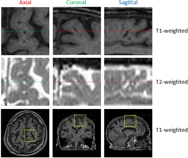
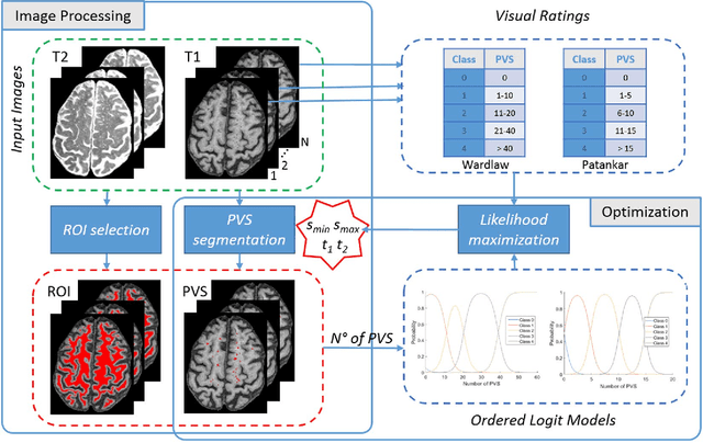
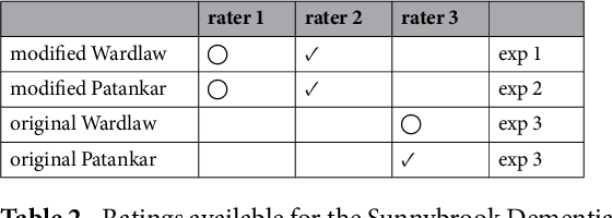
Abstract:Perivascular Spaces (PVS) are a recently recognised feature of Small Vessel Disease (SVD), also indicating neuroinflammation, and are an important part of the brain's circulation and glymphatic drainage system. Quantitative analysis of PVS on Magnetic Resonance Images (MRI) is important for understanding their relationship with neurological diseases. In this work, we propose a segmentation technique based on the 3D Frangi filtering for extraction of PVS from MRI. Based on prior knowledge from neuroradiological ratings of PVS, we used ordered logit models to optimise Frangi filter parameters in response to the variability in the scanner's parameters and study protocols. We optimized and validated our proposed models on two independent cohorts, a dementia sample (N=20) and patients who previously had mild to moderate stroke (N=48). Results demonstrate the robustness and generalisability of our segmentation method. Segmentation-based PVS burden estimates correlated with neuroradiological assessments (Spearman's $\rho$ = 0.74, p $<$ 0.001), suggesting the great potential of our proposed method
 Add to Chrome
 Add to Firefox
 Add to Edge