Amit Kharat
A Classical-Quantum Convolutional Neural Network for Detecting Pneumonia from Chest Radiographs
Feb 19, 2022


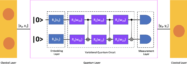
Abstract:While many quantum computing techniques for machine learning have been proposed, their performance on real-world datasets remains to be studied. In this paper, we explore how a variational quantum circuit could be integrated into a classical neural network for the problem of detecting pneumonia from chest radiographs. We substitute one layer of a classical convolutional neural network with a variational quantum circuit to create a hybrid neural network. We train both networks on an image dataset containing chest radiographs and benchmark their performance. To mitigate the influence of different sources of randomness in network training, we sample the results over multiple rounds. We show that the hybrid network outperforms the classical network on different performance measures, and that these improvements are statistically significant. Our work serves as an experimental demonstration of the potential of quantum computing to significantly improve neural network performance for real-world, non-trivial problems relevant to society and industry.
Levels of Autonomous Radiology
Dec 14, 2021Abstract:Radiology, being one of the younger disciplines of medicine with a history of just over a century, has witnessed tremendous technological advancements and has revolutionized the way we practice medicine today. In the last few decades, medical imaging modalities have generated seismic amounts of medical data. The development and adoption of Artificial Intelligence (AI) applications using this data will lead to the next phase of evolution in radiology. It will include automating laborious manual tasks such as annotations, report-generation, etc., along with the initial radiological assessment of cases to aid radiologists in their evaluation workflow. We propose a level-wise classification for the progression of automation in radiology, explaining AI assistance at each level with corresponding challenges and solutions. We hope that such discussions can help us address the challenges in a structured way and take the necessary steps to ensure the smooth adoption of new technologies in radiology.
Reducing Labelled Data Requirement for Pneumonia Segmentation using Image Augmentations
Feb 25, 2021

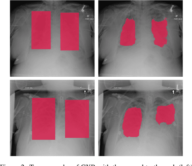
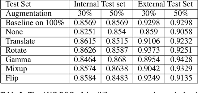
Abstract:Deep learning semantic segmentation algorithms can localise abnormalities or opacities from chest radiographs. However, the task of collecting and annotating training data is expensive and requires expertise which remains a bottleneck for algorithm performance. We investigate the effect of image augmentations on reducing the requirement of labelled data in the semantic segmentation of chest X-rays for pneumonia detection. We train fully convolutional network models on subsets of different sizes from the total training data. We apply a different image augmentation while training each model and compare it to the baseline trained on the entire dataset without augmentations. We find that rotate and mixup are the best augmentations amongst rotate, mixup, translate, gamma and horizontal flip, wherein they reduce the labelled data requirement by 70% while performing comparably to the baseline in terms of AUC and mean IoU in our experiments.
Key Technology Considerations in Developing and Deploying Machine Learning Models in Clinical Radiology Practice
Feb 03, 2021
Abstract:The use of machine learning to develop intelligent software tools for interpretation of radiology images has gained widespread attention in recent years. The development, deployment, and eventual adoption of these models in clinical practice, however, remains fraught with challenges. In this paper, we propose a list of key considerations that machine learning researchers must recognize and address to make their models accurate, robust, and usable in practice. Namely, we discuss: insufficient training data, decentralized datasets, high cost of annotations, ambiguous ground truth, imbalance in class representation, asymmetric misclassification costs, relevant performance metrics, generalization of models to unseen datasets, model decay, adversarial attacks, explainability, fairness and bias, and clinical validation. We describe each consideration and identify techniques to address it. Although these techniques have been discussed in prior research literature, by freshly examining them in the context of medical imaging and compiling them in the form of a laundry list, we hope to make them more accessible to researchers, software developers, radiologists, and other stakeholders.
Comparative Evaluation of 3D and 2D Deep Learning Techniques for Semantic Segmentation in CT Scans
Jan 19, 2021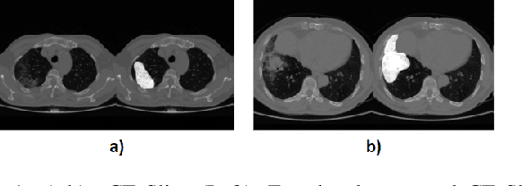

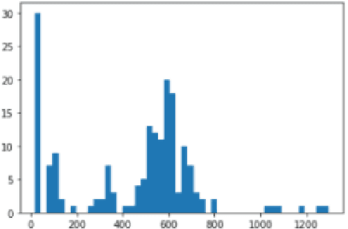
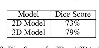
Abstract:Image segmentation plays a pivotal role in several medical-imaging applications by assisting the segmentation of the regions of interest. Deep learning-based approaches have been widely adopted for semantic segmentation of medical data. In recent years, in addition to 2D deep learning architectures, 3D architectures have been employed as the predictive algorithms for 3D medical image data. In this paper, we propose a 3D stack-based deep learning technique for segmenting manifestations of consolidation and ground-glass opacities in 3D Computed Tomography (CT) scans. We also present a comparison based on the segmentation results, the contextual information retained, and the inference time between this 3D technique and a traditional 2D deep learning technique. We also define the area-plot, which represents the peculiar pattern observed in the slice-wise areas of the pathology regions predicted by these deep learning models. In our exhaustive evaluation, 3D technique performs better than the 2D technique for the segmentation of CT scans. We get dice scores of 79% and 73% for the 3D and the 2D techniques respectively. The 3D technique results in a 5X reduction in the inference time compared to the 2D technique. Results also show that the area-plots predicted by the 3D model are more similar to the ground truth than those predicted by the 2D model. We also show how increasing the amount of contextual information retained during the training can improve the 3D model's performance.
Deep Learning Models for Calculation of Cardiothoracic Ratio from Chest Radiographs for Assisted Diagnosis of Cardiomegaly
Jan 19, 2021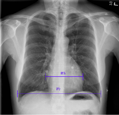

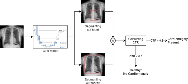
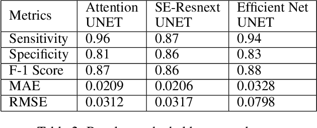
Abstract:We propose an automated method based on deep learning to compute the cardiothoracic ratio and detect the presence of cardiomegaly from chest radiographs. We develop two separate models to demarcate the heart and chest regions in an X-ray image using bounding boxes and use their outputs to calculate the cardiothoracic ratio. We obtain a sensitivity of 0.96 at a specificity of 0.81 with a mean absolute error of 0.0209 on a held-out test dataset and a sensitivity of 0.84 at a specificity of 0.97 with a mean absolute error of 0.018 on an independent dataset from a different hospital. We also compare three different segmentation model architectures for the proposed method and observe that Attention U-Net yields better results than SE-Resnext U-Net and EfficientNet U-Net. By providing a numeric measurement of the cardiothoracic ratio, we hope to mitigate human subjectivity arising out of visual assessment in the detection of cardiomegaly.
Automated Detection of COVID-19 from CT Scans Using Convolutional Neural Networks
Jun 23, 2020
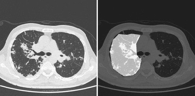
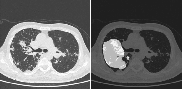

Abstract:COVID-19 is an infectious disease that causes respiratory problems similar to those caused by SARS-CoV (2003). Currently, swab samples are being used for its diagnosis. The most common testing method used is the RT-PCR method, which has high specificity but variable sensitivity. AI-based detection has the capability to overcome this drawback. In this paper, we propose a prospective method wherein we use chest CT scans to diagnose the patients for COVID-19 pneumonia. We use a set of open-source images, available as individual CT slices, and full CT scans from a private Indian Hospital to train our model. We build a 2D segmentation model using the U-Net architecture, which gives the output by marking out the region of infection. Our model achieves a sensitivity of 96.428% (95% CI: 88%-100%) and a specificity of 88.39% (95% CI: 82%-94%). Additionally, we derive a logic for converting our slice-level predictions to scan-level, which helps us reduce the false positives.
Role of Edge Device and Cloud Machine Learning in Point-of-Care Solutions Using Imaging Diagnostics for Population Screening
Jun 18, 2020
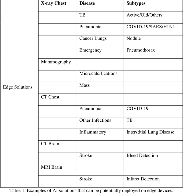

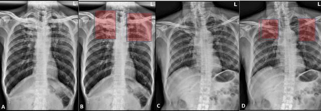
Abstract:Edge devices are revolutionizing diagnostics. Edge devices can reside within or adjacent to imaging tools such as digital Xray, CT, MRI, or ultrasound equipment. These devices are either CPUs or GPUs with advanced processing deep and machine learning (artificial intelligence) algorithms that assist in classification and triage solutions to flag studies as either normal or abnormal, TB or healthy (in case of TB screening), suspected COVID-19/other pneumonia or unremarkable (in hospital or hotspot settings). These can be deployed as screening point-of-care (PoC) solutions; this is particularly true for digital and portable X-ray devices. Edge device learning can also be used for mammography and CT studies where it can identify microcalcification and stroke, respectively. These solutions can be considered the first line of pre-screening before the imaging specialist actually reviews scans and makes a final diagnosis. The key advantage of these tools is that they are instant, can be deployed remotely where experts are not available to perform pre-screening before the experts actually review, and are not limited by internet bandwidth as the nano learning data centers are placed next to the device.
Automatic Grading of Knee Osteoarthritis on the Kellgren-Lawrence Scale from Radiographs Using Convolutional Neural Networks
Apr 18, 2020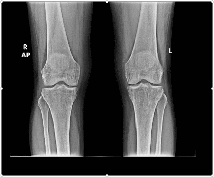

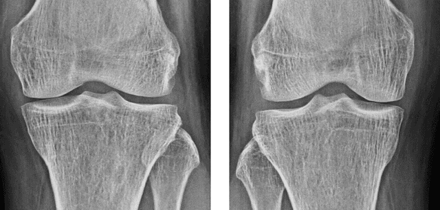
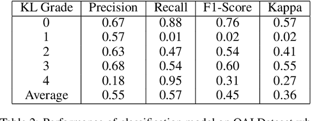
Abstract:The severity of knee osteoarthritis is graded using the 5-point Kellgren-Lawrence (KL) scale where healthy knees are assigned grade 0, and the subsequent grades 1-4 represent increasing severity of the affliction. Although several methods have been proposed in recent years to develop models that can automatically predict the KL grade from a given radiograph, most models have been developed and evaluated on datasets not sourced from India. These models fail to perform well on the radiographs of Indian patients. In this paper, we propose a novel method using convolutional neural networks to automatically grade knee radiographs on the KL scale. Our method works in two connected stages: in the first stage, an object detection model segments individual knees from the rest of the image; in the second stage, a regression model automatically grades each knee separately on the KL scale. We train our model using the publicly available Osteoarthritis Initiative (OAI) dataset and demonstrate that fine-tuning the model before evaluating it on a dataset from a private hospital significantly improves the mean absolute error from 1.09 (95% CI: 1.03-1.15) to 0.28 (95% CI: 0.25-0.32). Additionally, we compare classification and regression models built for the same task and demonstrate that regression outperforms classification.
 Add to Chrome
 Add to Firefox
 Add to Edge