Reducing Labelled Data Requirement for Pneumonia Segmentation using Image Augmentations
Paper and Code
Feb 25, 2021


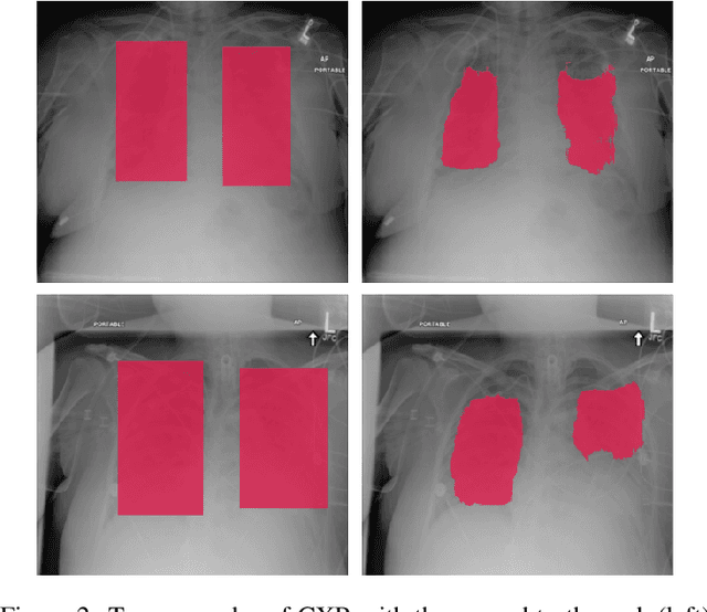
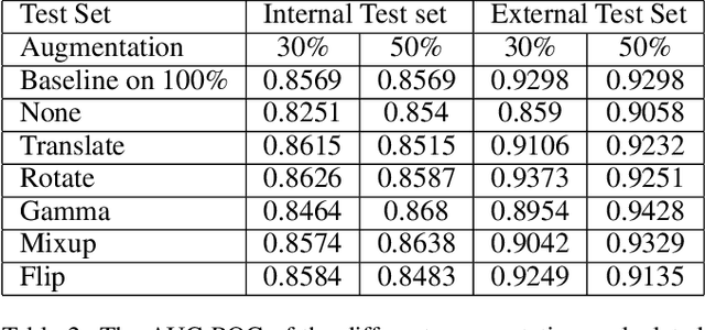
Deep learning semantic segmentation algorithms can localise abnormalities or opacities from chest radiographs. However, the task of collecting and annotating training data is expensive and requires expertise which remains a bottleneck for algorithm performance. We investigate the effect of image augmentations on reducing the requirement of labelled data in the semantic segmentation of chest X-rays for pneumonia detection. We train fully convolutional network models on subsets of different sizes from the total training data. We apply a different image augmentation while training each model and compare it to the baseline trained on the entire dataset without augmentations. We find that rotate and mixup are the best augmentations amongst rotate, mixup, translate, gamma and horizontal flip, wherein they reduce the labelled data requirement by 70% while performing comparably to the baseline in terms of AUC and mean IoU in our experiments.
 Add to Chrome
 Add to Firefox
 Add to Edge