Yuki Suzuki
Anatomy-aware Self-supervised Learning for Anomaly Detection in Chest Radiographs
May 09, 2022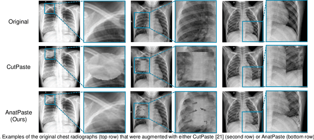
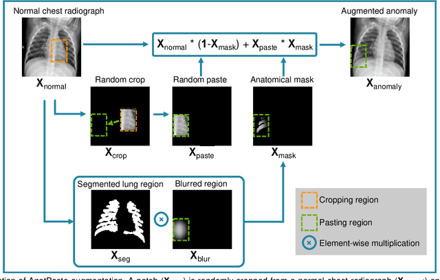
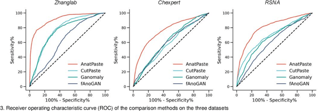
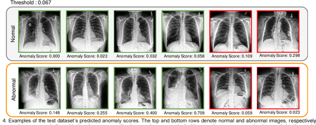
Abstract:Large numbers of labeled medical images are essential for the accurate detection of anomalies, but manual annotation is labor-intensive and time-consuming. Self-supervised learning (SSL) is a training method to learn data-specific features without manual annotation. Several SSL-based models have been employed in medical image anomaly detection. These SSL methods effectively learn representations in several field-specific images, such as natural and industrial product images. However, owing to the requirement of medical expertise, typical SSL-based models are inefficient in medical image anomaly detection. We present an SSL-based model that enables anatomical structure-based unsupervised anomaly detection (UAD). The model employs the anatomy-aware pasting (AnatPaste) augmentation tool. AnatPaste employs a threshold-based lung segmentation pretext task to create anomalies in normal chest radiographs, which are used for model pretraining. These anomalies are similar to real anomalies and help the model recognize them. We evaluate our model on three opensource chest radiograph datasets. Our model exhibit area under curves (AUC) of 92.1%, 78.7%, and 81.9%, which are the highest among existing UAD models. This is the first SSL model to employ anatomical information as a pretext task. AnatPaste can be applied in various deep learning models and downstream tasks. It can be employed for other modalities by fixing appropriate segmentation. Our code is publicly available at: https://github.com/jun-sato/AnatPaste.
Weak Supervision in Convolutional Neural Network for Semantic Segmentation of Diffuse Lung Diseases Using Partially Annotated Dataset
Mar 26, 2020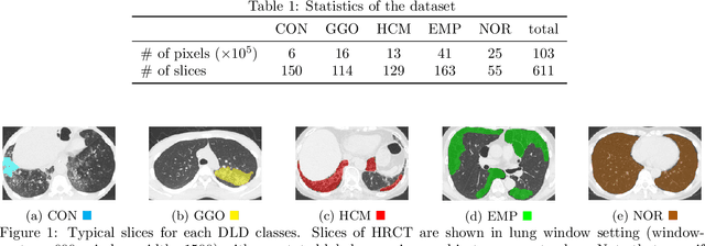
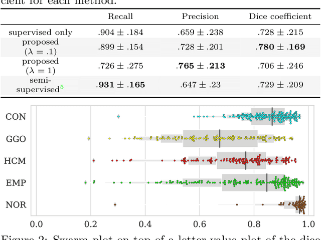
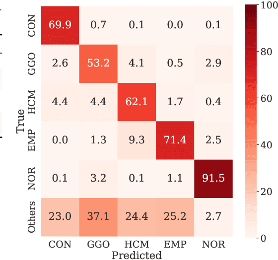
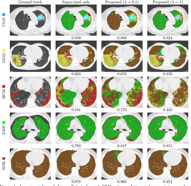
Abstract:Computer-aided diagnosis system for diffuse lung diseases (DLDs) is necessary for the objective assessment of the lung diseases. In this paper, we develop semantic segmentation model for 5 kinds of DLDs. DLDs considered in this work are consolidation, ground glass opacity, honeycombing, emphysema, and normal. Convolutional neural network (CNN) is one of the most promising technique for semantic segmentation among machine learning algorithms. While creating annotated dataset for semantic segmentation is laborious and time consuming, creating partially annotated dataset, in which only one chosen class is annotated for each image, is easier since annotators only need to focus on one class at a time during the annotation task. In this paper, we propose a new weak supervision technique that effectively utilizes partially annotated dataset. The experiments using partially annotated dataset composed 372 CT images demonstrated that our proposed technique significantly improved segmentation accuracy.
Automated Segmentation of Hip and Thigh Muscles in Metal Artifact-Contaminated CT using Convolutional Neural Network-Enhanced Normalized Metal Artifact Reduction
Jun 27, 2019


Abstract:In total hip arthroplasty, analysis of postoperative medical images is important to evaluate surgical outcome. Since Computed Tomography (CT) is most prevalent modality in orthopedic surgery, we aimed at the analysis of CT image. In this work, we focus on the metal artifact in postoperative CT caused by the metallic implant, which reduces the accuracy of segmentation especially in the vicinity of the implant. Our goal was to develop an automated segmentation method of the bones and muscles in the postoperative CT images. We propose a method that combines Normalized Metal Artifact Reduction (NMAR), which is one of the state-of-the-art metal artifact reduction methods, and a Convolutional Neural Network-based segmentation using two U-net architectures. The first U-net refines the result of NMAR and the muscle segmentation is performed by the second U-net. We conducted experiments using simulated images of 20 patients and real images of three patients to evaluate the segmentation accuracy of 19 muscles. In simulation study, the proposed method showed statistically significant improvement (p<0.05) in the average symmetric surface distance (ASD) metric for 14 muscles out of 19 muscles and the average ASD of all muscles from 1.17 +/- 0.543 mm (mean +/- std over all patients) to 1.10 +/- 0.509 mm over our previous method. The real image study using the manual trace of gluteus maximus and medius muscles showed ASD of 1.32 +/- 0.25 mm. Our future work includes training of a network in an end-to-end manner for both the metal artifact reduction and muscle segmentation.
 Add to Chrome
 Add to Firefox
 Add to Edge