Xinyun Zhong
Respiratory Subtraction for Pulmonary Microwave Ablation Evaluation
Aug 08, 2024


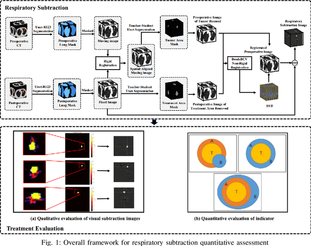
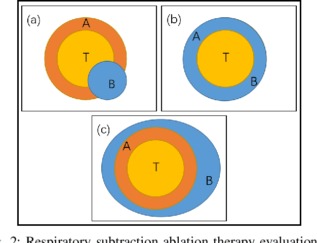
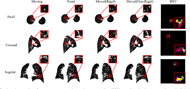
Abstract:Currently, lung cancer is a leading cause of global cancer mortality, often necessitating minimally invasive interventions. Microwave ablation (MWA) is extensively utilized for both primary and secondary lung tumors. Although numerous clinical guidelines and standards for MWA have been established, the clinical evaluation of ablation surgery remains challenging and requires long-term patient follow-up for confirmation. In this paper, we propose a method termed respiratory subtraction to evaluate lung tumor ablation therapy performance based on pre- and post-operative image guidance. Initially, preoperative images undergo coarse rigid registration to their corresponding postoperative positions, followed by further non-rigid registration. Subsequently, subtraction images are generated by subtracting the registered preoperative images from the postoperative ones. Furthermore, to enhance the clinical assessment of MWA treatment performance, we devise a quantitative analysis metric to evaluate ablation efficacy by comparing differences between tumor areas and treatment areas. To the best of our knowledge, this is the pioneering work in the field to facilitate the assessment of MWA surgery performance on pulmonary tumors. Extensive experiments involving 35 clinical cases further validate the efficacy of the respiratory subtraction method. The experimental results confirm the effectiveness of the respiratory subtraction method and the proposed quantitative evaluation metric in assessing lung tumor treatment.
PDS-MAR: a fine-grained Projection-Domain Segmentation-based Metal Artifact Reduction method for intraoperative CBCT images with guidewires
Jun 21, 2023Abstract:Since the invention of modern CT systems, metal artifacts have been a persistent problem. Due to increased scattering, amplified noise, and insufficient data collection, it is more difficult to suppress metal artifacts in cone-beam CT, limiting its use in human- and robot-assisted spine surgeries where metallic guidewires and screws are commonly used. In this paper, we demonstrate that conventional image-domain segmentation-based MAR methods are unable to eliminate metal artifacts for intraoperative CBCT images with guidewires. To solve this problem, we present a fine-grained projection-domain segmentation-based MAR method termed PDS-MAR, in which metal traces are augmented and segmented in the projection domain before being inpainted using triangular interpolation. In addition, a metal reconstruction phase is proposed to restore metal areas in the image domain. The digital phantom study and real CBCT data study demonstrate that the proposed algorithm achieves significantly better artifact suppression than other comparing methods and has the potential to advance the use of intraoperative CBCT imaging in clinical spine surgeries.
 Add to Chrome
 Add to Firefox
 Add to Edge