Tomasz Włodarczyk
FetalNet: Multi-task deep learning framework for fetal ultrasound biometric measurements
Jul 14, 2021

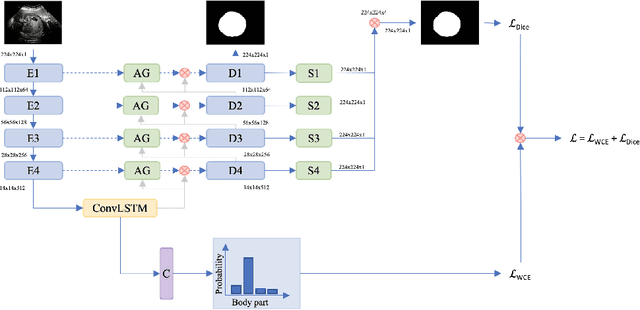
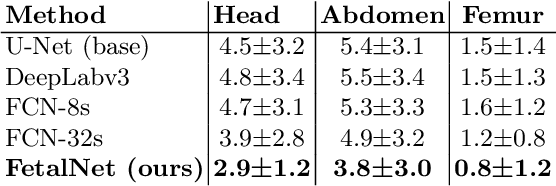
Abstract:In this paper, we propose an end-to-end multi-task neural network called FetalNet with an attention mechanism and stacked module for spatio-temporal fetal ultrasound scan video analysis. Fetal biometric measurement is a standard examination during pregnancy used for the fetus growth monitoring and estimation of gestational age and fetal weight. The main goal in fetal ultrasound scan video analysis is to find proper standard planes to measure the fetal head, abdomen and femur. Due to natural high speckle noise and shadows in ultrasound data, medical expertise and sonographic experience are required to find the appropriate acquisition plane and perform accurate measurements of the fetus. In addition, existing computer-aided methods for fetal US biometric measurement address only one single image frame without considering temporal features. To address these shortcomings, we propose an end-to-end multi-task neural network for spatio-temporal ultrasound scan video analysis to simultaneously localize, classify and measure the fetal body parts. We propose a new encoder-decoder segmentation architecture that incorporates a classification branch. Additionally, we employ an attention mechanism with a stacked module to learn salient maps to suppress irrelevant US regions and efficient scan plane localization. We trained on the fetal ultrasound video comes from routine examinations of 700 different patients. Our method called FetalNet outperforms existing state-of-the-art methods in both classification and segmentation in fetal ultrasound video recordings.
Convolutional Neural Networks in Orthodontics: a review
Apr 18, 2021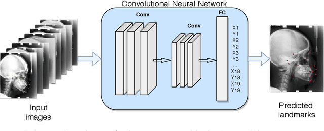
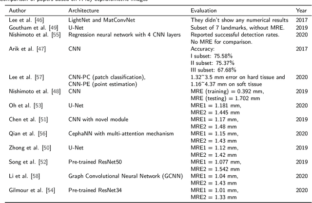
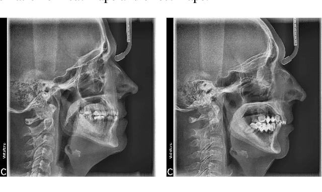
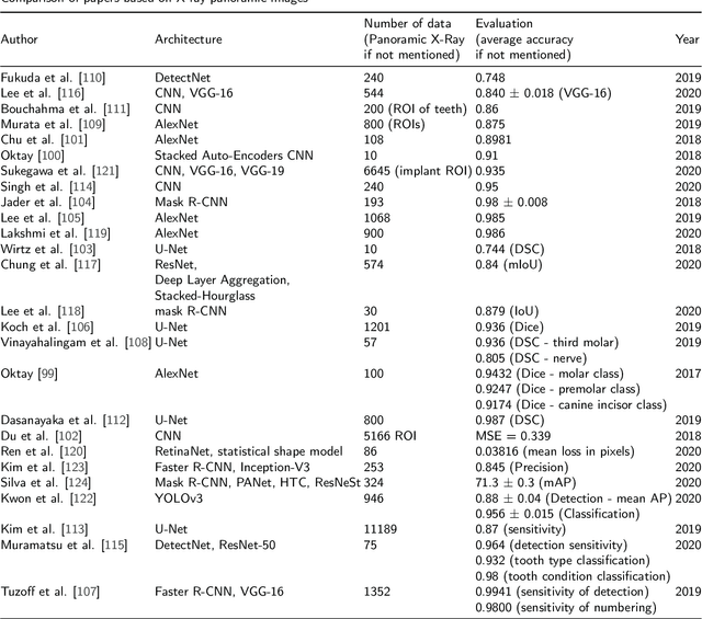
Abstract:Convolutional neural networks (CNNs) are used in many areas of computer vision, such as object tracking and recognition, security, military, and biomedical image analysis. This review presents the application of convolutional neural networks in one of the fields of dentistry - orthodontics. Advances in medical imaging technologies and methods allow CNNs to be used in orthodontics to shorten the planning time of orthodontic treatment, including an automatic search of landmarks on cephalometric X-ray images, tooth segmentation on Cone-Beam Computed Tomography (CBCT) images or digital models, and classification of defects on X-Ray panoramic images. In this work, we describe the current methods, the architectures of deep convolutional neural networks used, and their implementations, together with a comparison of the results achieved by them. The promising results and visualizations of the described studies show that the use of methods based on convolutional neural networks allows for the improvement of computer-based orthodontic treatment planning, both by reducing the examination time and, in many cases, by performing the analysis much more accurately than a manual orthodontist does.
Spontaneous preterm birth prediction using convolutional neural networks
Aug 21, 2020
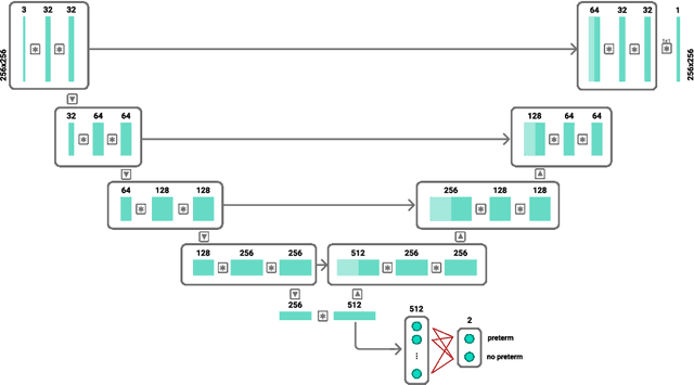
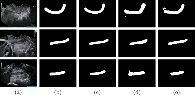
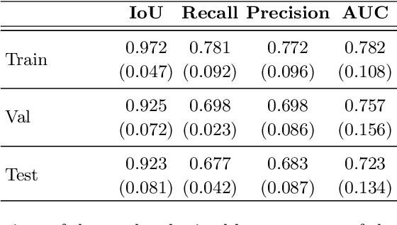
Abstract:An estimated 15 million babies are born too early every year. Approximately 1 million children die each year due to complications of preterm birth (PTB). Many survivors face a lifetime of disability, including learning disabilities and visual and hearing problems. Although manual analysis of ultrasound images (US) is still prevalent, it is prone to errors due to its subjective component and complex variations in the shape and position of organs across patients. In this work, we introduce a conceptually simple convolutional neural network (CNN) trained for segmenting prenatal ultrasound images and classifying task for the purpose of preterm birth detection. Our method efficiently segments different types of cervixes in transvaginal ultrasound images while simultaneously predicting a preterm birth based on extracted image features without human oversight. We employed three popular network models: U-Net, Fully Convolutional Network, and Deeplabv3 for the cervix segmentation task. Based on the conducted results and model efficiency, we decided to extend U-Net by adding a parallel branch for classification task. The proposed model is trained and evaluated on a dataset consisting of 354 2D transvaginal ultrasound images and achieved a segmentation accuracy with a mean Jaccard coefficient index of 0.923 $\pm$ 0.081 and a classification sensitivity of 0.677 $\pm$ 0.042 with a 3.49\% false positive rate. Our method obtained better results in the prediction of preterm birth based on transvaginal ultrasound images compared to state-of-the-art methods.
Estimation of preterm birth markers with U-Net segmentation network
Aug 24, 2019
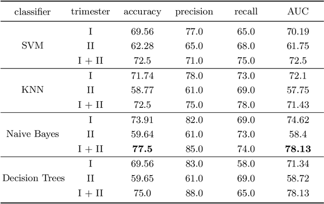

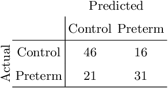
Abstract:Preterm birth is the most common cause of neonatal death. Current diagnostic methods that assess the risk of preterm birth involve the collection of maternal characteristics and transvaginal ultrasound imaging conducted in the first and second trimester of pregnancy. Analysis of the ultrasound data is based on visual inspection of images by gynaecologist, sometimes supported by hand-designed image features such as cervical length. Due to the complexity of this process and its subjective component, approximately 30% of spontaneous preterm deliveries are not correctly predicted. Moreover, 10% of the predicted preterm deliveries are false-positives. In this paper, we address the problem of predicting spontaneous preterm delivery using machine learning. To achieve this goal, we propose to first use a deep neural network architecture for segmenting prenatal ultrasound images and then automatically extract two biophysical ultrasound markers, cervical length (CL) and anterior cervical angle (ACA), from the resulting images. Our method allows to estimate ultrasound markers without human oversight. Furthermore, we show that CL and ACA markers, when combined, allow us to decrease false-negative ratio from 30% to 18%. Finally, contrary to the current approaches to diagnostics methods that rely only on gynaecologist's expertise, our method introduce objectively obtained results.
 Add to Chrome
 Add to Firefox
 Add to Edge