Titas De
A Wearable Social Interaction Aid for Children with Autism
Apr 19, 2020
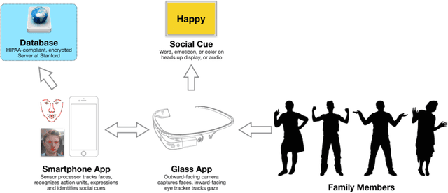
Abstract:With most recent estimates giving an incidence rate of 1 in 68 children in the United States, the autism spectrum disorder (ASD) is a growing public health crisis. Many of these children struggle to make eye contact, recognize facial expressions, and engage in social interactions. Today the standard for treatment of the core autism-related deficits focuses on a form of behavior training known as Applied Behavioral Analysis. To address perceived deficits in expression recognition, ABA approaches routinely involve the use of prompts such as flash cards for repetitive emotion recognition training via memorization. These techniques must be administered by trained practitioners and often at clinical centers that are far outnumbered by and out of reach from the many children and families in need of attention. Waitlists for access are up to 18 months long, and this wait may lead to children regressing down a path of isolation that worsens their long-term prognosis. There is an urgent need to innovate new methods of care delivery that can appropriately empower caregivers of children at risk or with a diagnosis of autism, and that capitalize on mobile tools and wearable devices for use outside of clinical settings.
Introducing Anisotropic Minkowski Functionals for Local Structure Analysis and Prediction of Biomechanical Strength of Proximal Femur Specimens
Apr 02, 2020


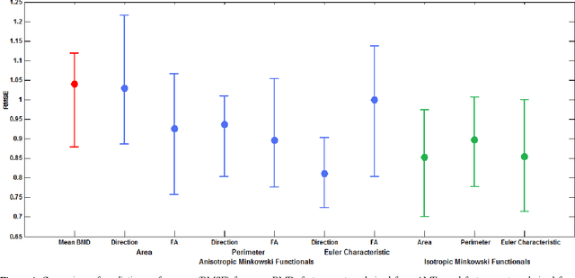
Abstract:Bone fragility and fracture caused by osteoporosis or injury are prevalent in adults over the age of 50 and can reduce their quality of life. Hence, predicting the biomechanical bone strength, specifically of the proximal femur, through non-invasive imaging-based methods is an important goal for the diagnosis of Osteoporosis as well as estimating fracture risk. Dual X-ray absorptiometry (DXA) has been used as a standard clinical procedure for assessment and diagnosis of bone strength and osteoporosis through bone mineral density (BMD) measurements. However, previous studies have shown that quantitative computer tomography (QCT) can be more sensitive and specific to trabecular bone characterization because it reduces the overlap effects and interferences from the surrounding soft tissue and cortical shell. This study proposes a new method to predict the bone strength of proximal femur specimens from quantitative multi-detector computer tomography (MDCT) images. Texture analysis methods such as conventional statistical moments (BMD mean), Isotropic Minkowski Functionals (IMF) and Anisotropic Minkowski Functionals (AMF) are used to quantify BMD properties of the trabecular bone micro-architecture. Combinations of these extracted features are then used to predict the biomechanical strength of the femur specimens using sophisticated machine learning techniques such as multiregression (MultiReg) and support vector regression with linear kernel (SVRlin). The prediction performance achieved with these feature sets is compared to the standard approach that uses the mean BMD of the specimens and multiregression models using root mean square error (RMSE).
Introducing Anisotropic Minkowski Functionals and Quantitative Anisotropy Measures for Local Structure Analysis in Biomedical Imaging
Apr 02, 2020


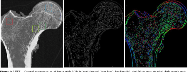
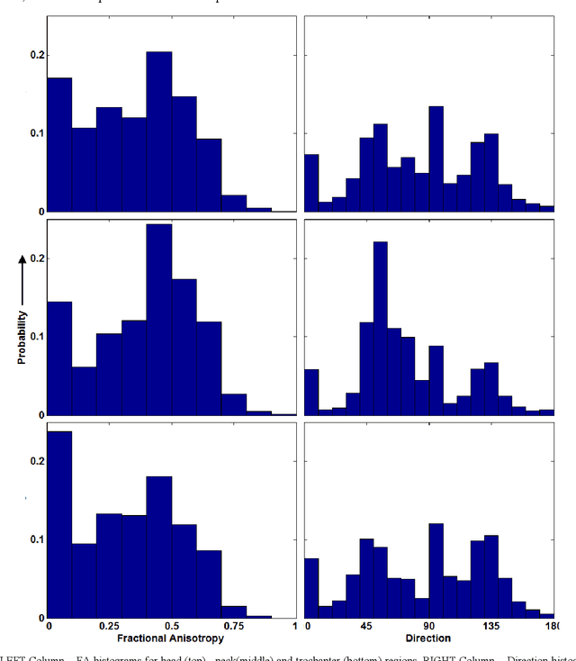
Abstract:The ability of Minkowski Functionals to characterize local structure in different biological tissue types has been demonstrated in a variety of medical image processing tasks. We introduce anisotropic Minkowski Functionals (AMFs) as a novel variant that captures the inherent anisotropy of the underlying gray-level structures. To quantify the anisotropy characterized by our approach, we further introduce a method to compute a quantitative measure motivated by a technique utilized in MR diffusion tensor imaging, namely fractional anisotropy. We showcase the applicability of our method in the research context of characterizing the local structure properties of trabecular bone micro-architecture in the proximal femur as visualized on multi-detector CT. To this end, AMFs were computed locally for each pixel of ROIs extracted from the head, neck and trochanter regions. Fractional anisotropy was then used to quantify the local anisotropy of the trabecular structures found in these ROIs and to compare its distribution in different anatomical regions. Our results suggest a significantly greater concentration of anisotropic trabecular structures in the head and neck regions when compared to the trochanter region (p < 10-4). We also evaluated the ability of such AMFs to predict bone strength in the femoral head of proximal femur specimens obtained from 50 donors. Our results suggest that such AMFs, when used in conjunction with multi-regression models, can outperform more conventional features such as BMD in predicting failure load. We conclude that such anisotropic Minkowski Functionals can capture valuable information regarding directional attributes of local structure, which may be useful in a wide scope of biomedical imaging applications.
 Add to Chrome
 Add to Firefox
 Add to Edge