Axel Wismueller
Enhancing Graph Attention Neural Network Performance for Marijuana Consumption Classification through Large-scale Augmented Granger Causality (lsAGC) Analysis of Functional MR Images
Oct 24, 2024Abstract:In the present research, the effectiveness of large-scale Augmented Granger Causality (lsAGC) as a tool for gauging brain network connectivity was examined to differentiate between marijuana users and typical controls by utilizing resting-state functional Magnetic Resonance Imaging (fMRI). The relationship between marijuana consumption and alterations in brain network connectivity is a recognized fact in scientific literature. This study probes how lsAGC can accurately discern these changes. The technique used integrates dimension reduction with the augmentation of source time-series in a model that predicts time-series, which helps in estimating the directed causal relationships among fMRI time-series. As a multivariate approach, lsAGC uncovers the connection of the inherent dynamic system while considering all other time-series. A dataset of 60 adults with an ADHD diagnosis during childhood, drawn from the Addiction Connectome Preprocessed Initiative (ACPI), was used in the study. The brain connections assessed by lsAGC were utilized as classification attributes. A Graph Attention Neural Network (GAT) was chosen to carry out the classification task, particularly for its ability to harness graph-based data and recognize intricate interactions between brain regions, making it appropriate for fMRI-based brain connectivity data. The performance was analyzed using a five-fold cross-validation system. The average accuracy achieved by the correlation coefficient method was roughly 52.98%, with a 1.65 standard deviation, whereas the lsAGC approach yielded an average accuracy of 61.47%, with a standard deviation of 1.44. The suggested method enhances the body of knowledge in the field of neuroimaging-based classification and emphasizes the necessity to consider directed causal connections in brain network connectivity analysis when studying marijuana's effects on the brain.
Cross Modal Global Local Representation Learning from Radiology Reports and X-Ray Chest Images
Jan 26, 2023Abstract:Deep learning models can be applied successfully in real-work problems; however, training most of these models requires massive data. Recent methods use language and vision, but unfortunately, they rely on datasets that are not usually publicly available. Here we pave the way for further research in the multimodal language-vision domain for radiology. In this paper, we train a representation learning method that uses local and global representations of the language and vision through an attention mechanism and based on the publicly available Indiana University Radiology Report (IU-RR) dataset. Furthermore, we use the learned representations to diagnose five lung pathologies: atelectasis, cardiomegaly, edema, pleural effusion, and consolidation. Finally, we use both supervised and zero-shot classifications to extensively analyze the performance of the representation learning on the IU-RR dataset. Average Area Under the Curve (AUC) is used to evaluate the accuracy of the classifiers for classifying the five lung pathologies. The average AUC for classifying the five lung pathologies on the IU-RR test set ranged from 0.85 to 0.87 using the different training datasets, namely CheXpert and CheXphoto. These results compare favorably to other studies using UI-RR. Extensive experiments confirm consistent results for classifying lung pathologies using the multimodal global local representations of language and vision information.
Re-defining Radiology Quality Assurance (QA) -- Artificial Intelligence (AI)-Based QA by Restricted Investigation of Unequal Scores (AQUARIUS)
May 03, 2022Abstract:There is an urgent need for streamlining radiology Quality Assurance (QA) programs to make them better and faster. Here, we present a novel approach, Artificial Intelligence (AI)-Based QUality Assurance by Restricted Investigation of Unequal Scores (AQUARIUS), for re-defining radiology QA, which reduces human effort by up to several orders of magnitude over existing approaches. AQUARIUS typically includes automatic comparison of AI-based image analysis with natural language processing (NLP) on radiology reports. Only the usually small subset of cases with discordant reads is subsequently reviewed by human experts. To demonstrate the clinical applicability of AQUARIUS, we performed a clinical QA study on Intracranial Hemorrhage (ICH) detection in 1936 head CT scans from a large academic hospital. Immediately following image acquisition, scans were automatically analyzed for ICH using a commercially available software (Aidoc, Tel Aviv, Israel). Cases rated positive for ICH by AI (ICH-AI+) were automatically flagged in radiologists' reading worklists, where flagging was randomly switched off with probability 50%. Using AQUARIUS with NLP on final radiology reports and targeted expert neuroradiology review of only 29 discordantly classified cases reduced the human QA effort by 98.5%, where we found a total of six non-reported true ICH+ cases, with radiologists' missed ICH detection rates of 0.52% and 2.5% for flagged and non-flagged cases, respectively. We conclude that AQUARIUS, by combining AI-based image analysis with NLP-based pre-selection of cases for targeted human expert review, can efficiently identify missed findings in radiology studies and significantly expedite radiology QA programs in a hybrid human-machine interoperability approach.
Introducing Anisotropic Minkowski Functionals and Quantitative Anisotropy Measures for Local Structure Analysis in Biomedical Imaging
Apr 02, 2020


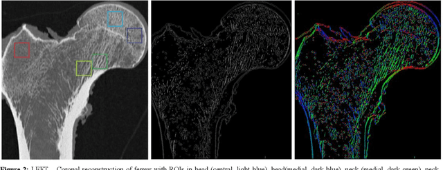
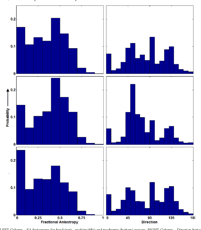
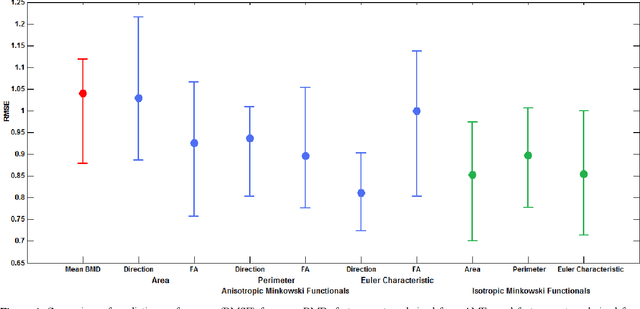
Abstract:The ability of Minkowski Functionals to characterize local structure in different biological tissue types has been demonstrated in a variety of medical image processing tasks. We introduce anisotropic Minkowski Functionals (AMFs) as a novel variant that captures the inherent anisotropy of the underlying gray-level structures. To quantify the anisotropy characterized by our approach, we further introduce a method to compute a quantitative measure motivated by a technique utilized in MR diffusion tensor imaging, namely fractional anisotropy. We showcase the applicability of our method in the research context of characterizing the local structure properties of trabecular bone micro-architecture in the proximal femur as visualized on multi-detector CT. To this end, AMFs were computed locally for each pixel of ROIs extracted from the head, neck and trochanter regions. Fractional anisotropy was then used to quantify the local anisotropy of the trabecular structures found in these ROIs and to compare its distribution in different anatomical regions. Our results suggest a significantly greater concentration of anisotropic trabecular structures in the head and neck regions when compared to the trochanter region (p < 10-4). We also evaluated the ability of such AMFs to predict bone strength in the femoral head of proximal femur specimens obtained from 50 donors. Our results suggest that such AMFs, when used in conjunction with multi-regression models, can outperform more conventional features such as BMD in predicting failure load. We conclude that such anisotropic Minkowski Functionals can capture valuable information regarding directional attributes of local structure, which may be useful in a wide scope of biomedical imaging applications.
 Add to Chrome
 Add to Firefox
 Add to Edge