 Add to Chrome
 Add to Firefox
 Add to Edge
TCIP: Threshold-Controlled Iterative Pyramid Network for Deformable Medical Image Registration
Oct 09, 2025Although pyramid networks have demonstrated superior performance in deformable medical image registration, their decoder architectures are inherently prone to propagating and accumulating anatomical structure misalignments. Moreover, most existing models do not adaptively determine the number of iterations for optimization under varying deformation requirements across images, resulting in either premature termination or excessive iterations that degrades registration accuracy. To effectively mitigate the accumulation of anatomical misalignments, we propose the Feature-Enhanced Residual Module (FERM) as the core component of each decoding layer in the pyramid network. FERM comprises three sequential blocks that extract anatomical semantic features, learn to suppress irrelevant features, and estimate the final deformation field, respectively. To adaptively determine the number of iterations for varying images, we propose the dual-stage Threshold-Controlled Iterative (TCI) strategy. In the first stage, TCI assesses registration stability and with asserted stability, it continues with the second stage to evaluate convergence. We coin the model that integrates FERM and TCI as Threshold-Controlled Iterative Pyramid (TCIP). Extensive experiments on three public brain MRI datasets and one abdomen CT dataset demonstrate that TCIP outperforms the state-of-the-art (SOTA) registration networks in terms of accuracy, while maintaining comparable inference speed and a compact model parameter size. Finally, we assess the generalizability of FERM and TCI by integrating them with existing registration networks and further conduct ablation studies to validate the effectiveness of these two proposed methods.
UniReg: Foundation Model for Controllable Medical Image Registration
Mar 17, 2025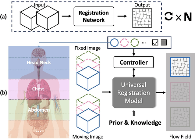
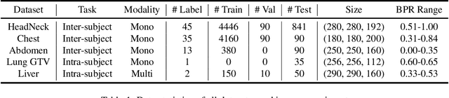
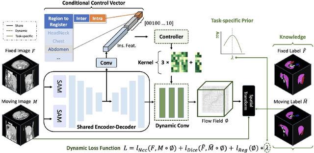
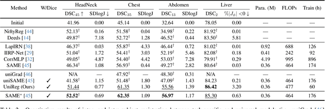
Learning-based medical image registration has achieved performance parity with conventional methods while demonstrating a substantial advantage in computational efficiency. However, learning-based registration approaches lack generalizability across diverse clinical scenarios, requiring the laborious development of multiple isolated networks for specific registration tasks, e.g., inter-/intra-subject registration or organ-specific alignment. % To overcome this limitation, we propose \textbf{UniReg}, the first interactive foundation model for medical image registration, which combines the precision advantages of task-specific learning methods with the generalization of traditional optimization methods. Our key innovation is a unified framework for diverse registration scenarios, achieved through a conditional deformation field estimation within a unified registration model. This is realized through a dynamic learning paradigm that explicitly encodes: (1) anatomical structure priors, (2) registration type constraints (inter/intra-subject), and (3) instance-specific features, enabling the generation of scenario-optimal deformation fields. % Through comprehensive experiments encompassing $90$ anatomical structures at different body regions, our UniReg model demonstrates comparable performance with contemporary state-of-the-art methodologies while achieving ~50\% reduction in required training iterations relative to the conventional learning-based paradigm. This optimization contributes to a significant reduction in computational resources, such as training time. Code and model will be available.
Leveraging Semantic Asymmetry for Precise Gross Tumor Volume Segmentation of Nasopharyngeal Carcinoma in Planning CT
Nov 27, 2024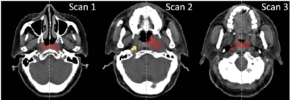
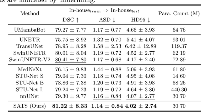
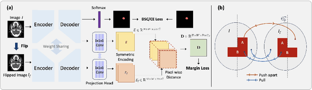
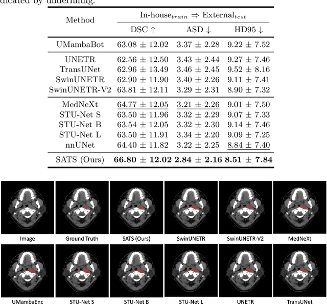
In the radiation therapy of nasopharyngeal carcinoma (NPC), clinicians typically delineate the gross tumor volume (GTV) using non-contrast planning computed tomography to ensure accurate radiation dose delivery. However, the low contrast between tumors and adjacent normal tissues necessitates that radiation oncologists manually delineate the tumors, often relying on diagnostic MRI for guidance. % In this study, we propose a novel approach to directly segment NPC gross tumors on non-contrast planning CT images, circumventing potential registration errors when aligning MRI or MRI-derived tumor masks to planning CT. To address the low contrast issues between tumors and adjacent normal structures in planning CT, we introduce a 3D Semantic Asymmetry Tumor segmentation (SATs) method. Specifically, we posit that a healthy nasopharyngeal region is characteristically bilaterally symmetric, whereas the emergence of nasopharyngeal carcinoma disrupts this symmetry. Then, we propose a Siamese contrastive learning segmentation framework that minimizes the voxel-wise distance between original and flipped areas without tumor and encourages a larger distance between original and flipped areas with tumor. Thus, our approach enhances the sensitivity of features to semantic asymmetries. % Extensive experiments demonstrate that the proposed SATs achieves the leading NPC GTV segmentation performance in both internal and external testing, \emph{e.g.}, with at least 2\% absolute Dice score improvement and 12\% average distance error reduction when compared to other state-of-the-art methods in the external testing.