Subhamoy Mandal
RAA-MIL: A Novel Framework for Classification of Oral Cytology
Nov 15, 2025Abstract:Cytology is a valuable tool for early detection of oral squamous cell carcinoma (OSCC). However, manual examination of cytology whole slide images (WSIs) is slow, subjective, and depends heavily on expert pathologists. To address this, we introduce the first weakly supervised deep learning framework for patient-level diagnosis of oral cytology whole slide images, leveraging the newly released Oral Cytology Dataset [1], which provides annotated cytology WSIs from ten medical centres across India. Each patient case is represented as a bag of cytology patches and assigned a diagnosis label (Healthy, Benign, Oral Potentially Malignant Disorders (OPMD), OSCC) by an in-house expert pathologist. These patient-level weak labels form a new extension to the dataset. We evaluate a baseline multiple-instance learning (MIL) model and a proposed Region-Affinity Attention MIL (RAA-MIL) that models spatial relationships between regions within each slide. The RAA-MIL achieves an average accuracy of 72.7%, weighted F1-score of 0.69 on an unseen test set, outperforming the baseline. This study establishes the first patient-level weakly supervised benchmark for oral cytology and moves toward reliable AI-assisted digital pathology.
Multimodal RGB-HSI Feature Fusion with Patient-Aware Incremental Heuristic Meta-Learning for Oral Lesion Classification
Nov 15, 2025


Abstract:Early detection of oral cancer and potentially malignant disorders is challenging in low-resource settings due to limited annotated data. We present a unified four-class oral lesion classifier that integrates deep RGB embeddings, hyperspectral reconstruction, handcrafted spectral-textural descriptors, and demographic metadata. A pathologist-verified subset of oral cavity images was curated and processed using a fine-tuned ConvNeXt-v2 encoder, followed by RGB-to-HSI reconstruction into 31-band hyperspectral cubes. Haemoglobin-sensitive indices, texture features, and spectral-shape measures were extracted and fused with deep and clinical features. Multiple machine-learning models were assessed with patient-wise validation. We further introduce an incremental heuristic meta-learner (IHML) that combines calibrated base classifiers through probabilistic stacking and patient-level posterior smoothing. On an unseen patient split, the proposed framework achieved a macro F1 of 66.23% and an accuracy of 64.56%. Results demonstrate that hyperspectral reconstruction and uncertainty-aware meta-learning substantially improve robustness for real-world oral lesion screening.
Multiscale Color Guided Attention Ensemble Classifier for Age-Related Macular Degeneration using Concurrent Fundus and Optical Coherence Tomography Images
Sep 01, 2024


Abstract:Automatic diagnosis techniques have evolved to identify age-related macular degeneration (AMD) by employing single modality Fundus images or optical coherence tomography (OCT). To classify ocular diseases, fundus and OCT images are the most crucial imaging modalities used in the clinical setting. Most deep learning-based techniques are established on a single imaging modality, which contemplates the ocular disorders to a specific extent and disregards other modality that comprises exhaustive information among distinct imaging modalities. This paper proposes a modality-specific multiscale color space embedding integrated with the attention mechanism based on transfer learning for classification (MCGAEc), which can efficiently extract the distinct modality information at various scales using the distinct color spaces. In this work, we first introduce the modality-specific multiscale color space encoder model, which includes diverse feature representations by integrating distinct characteristic color spaces on a multiscale into a unified framework. The extracted features from the prior encoder module are incorporated with the attention mechanism to extract the global features representation, which is integrated with the prior extracted features and transferred to the random forest classifier for the classification of AMD. To analyze the performance of the proposed MCGAEc method, a publicly available multi-modality dataset from Project Macula for AMD is utilized and compared with the existing models.
Speckle Noise Reduction in Ultrasound Images using Denoising Auto-encoder with Skip Connection
Mar 05, 2024


Abstract:Ultrasound is a widely used medical tool for non-invasive diagnosis, but its images often contain speckle noise which can lower their resolution and contrast-to-noise ratio. This can make it more difficult to extract, recognize, and analyze features in the images, as well as impair the accuracy of computer-assisted diagnostic techniques and the ability of doctors to interpret the images. Reducing speckle noise, therefore, is a crucial step in the preprocessing of ultrasound images. Researchers have proposed several speckle reduction methods, but no single method takes all relevant factors into account. In this paper, we compare seven such methods: Median, Gaussian, Bilateral, Average, Weiner, Anisotropic and Denoising auto-encoder without and with skip connections in terms of their ability to preserve features and edges while effectively reducing noise. In an experimental study, a convolutional noise-removing auto-encoder with skip connection, a deep learning method, was used to improve ultrasound images of breast cancer. This method involved adding speckle noise at various levels. The results of the deep learning method were compared to those of traditional image enhancement methods, and it was found that the proposed method was more effective. To assess the performance of these algorithms, we use three established evaluation metrics and present both filtered images and statistical data.
Deep Learning based Skin-layer Segmentation for Characterizing Cutaneous Wounds from Optical Coherence Tomography Images
Jun 02, 2023


Abstract:Optical coherence tomography (OCT) is a medical imaging modality that allows us to probe deeper substructures of skin. The state-of-the-art wound care prediction and monitoring methods are based on visual evaluation and focus on surface information. However, research studies have shown that sub-surface information of the wound is critical for understanding the wound healing progression. This work demonstrated the use of OCT as an effective imaging tool for objective and non-invasive assessments of wound severity, the potential for healing, and healing progress by measuring the optical characteristics of skin components. We have demonstrated the efficacy of OCT in studying wound healing progress in vivo small animal models. Automated analysis of OCT datasets poses multiple challenges, such as limitations in the training dataset size, variation in data distribution induced by uncertainties in sample quality and experiment conditions. We have employed a U-Net-based model for the segmentation of skin layers based on OCT images and to study epithelial and regenerated tissue thickness wound closure dynamics and thus quantify the progression of wound healing. In the experimental evaluation of the OCT skin image datasets, we achieved the objective of skin layer segmentation with an average intersection over union (IOU) of 0.9234. The results have been corroborated using gold-standard histology images and co-validated using inputs from pathologists. Clinical Relevance: To monitor wound healing progression without disrupting the healing procedure by superficial, noninvasive means via the identification of pixel characteristics of individual layers.
* Accepted
Maximum entropy based non-negative optoacoustic tomographic image reconstruction
Jul 26, 2017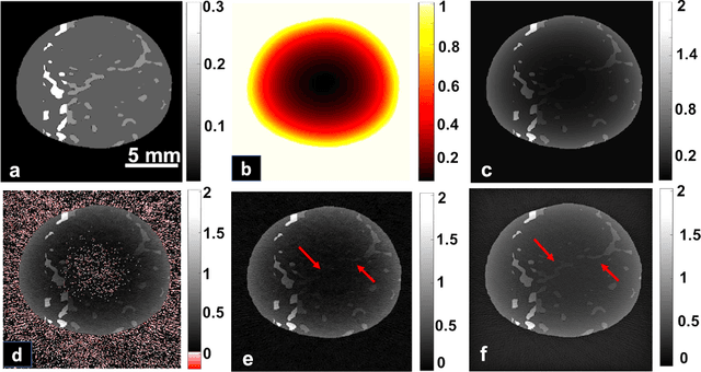
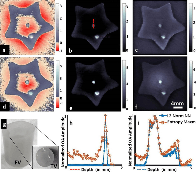

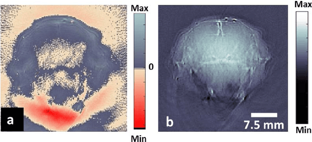
Abstract:Optoacoustic (photoacoustic) tomography reconstructs maps of the initial pressure rise induced by the absorption of light pulses in tissue. In practice, due to inaccurate assumptions in the forward model employed, noise and other experimental factors, the images often contain errors, occasionally manifested as negative values. We present optoacoustic tomography based on an entropy maximization algorithm that uses logarithmic regularization as a potent method for imparting non-negative image reconstruction. We experimentally investigate the performance achieved by the entropy maximization scheme on phantoms and in vivo samples. The findings demonstrate that the proposed scheme reconstructs physically relevant image values devoid of unwanted negative contrast, thus improving quantitative imaging performance.
Visual Quality Enhancement in Optoacoustic Tomography using Active Contour Segmentation Priors
Apr 11, 2016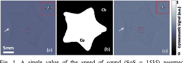
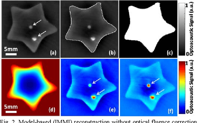
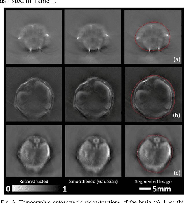
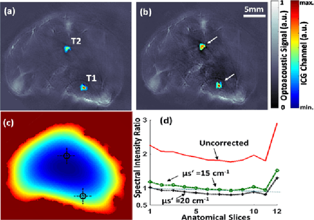
Abstract:Segmentation of biomedical images is essential for studying and characterizing anatomical structures, detection and evaluation of pathological tissues. Segmentation has been further shown to enhance the reconstruction performance in many tomographic imaging modalities by accounting for heterogeneities of the excitation field and tissue properties in the imaged region. This is particularly relevant in optoacoustic tomography, where discontinuities in the optical and acoustic tissue properties, if not properly accounted for, may result in deterioration of the imaging performance. Efficient segmentation of optoacoustic images is often hampered by the relatively low intrinsic contrast of large anatomical structures, which is further impaired by the limited angular coverage of some commonly employed tomographic imaging configurations. Herein, we analyze the performance of active contour models for boundary segmentation in cross-sectional optoacoustic tomography. The segmented mask is employed to construct a two compartment model for the acoustic and optical parameters of the imaged tissues, which is subsequently used to improve accuracy of the image reconstruction routines. The performance of the suggested segmentation and modeling approach are showcased in tissue-mimicking phantoms and small animal imaging experiments.
Grading of Mammalian Cumulus Oocyte Complexes using Machine Learning for in Vitro Embryo Culture
Mar 05, 2016


Abstract:Visual observation of Cumulus Oocyte Complexes provides only limited information about its functional competence, whereas the molecular evaluations methods are cumbersome or costly. Image analysis of mammalian oocytes can provide attractive alternative to address this challenge. However, it is complex, given the huge number of oocytes under inspection and the subjective nature of the features inspected for identification. Supervised machine learning methods like random forest with annotations from expert biologists can make the analysis task standardized and reduces inter-subject variability. We present a semi-automatic framework for predicting the class an oocyte belongs to, based on multi-object parametric segmentation on the acquired microscopic image followed by a feature based classification using random forests.
Multiscale edge detection and parametric shape modeling for boundary delineation in optoacoustic images
Jun 09, 2015

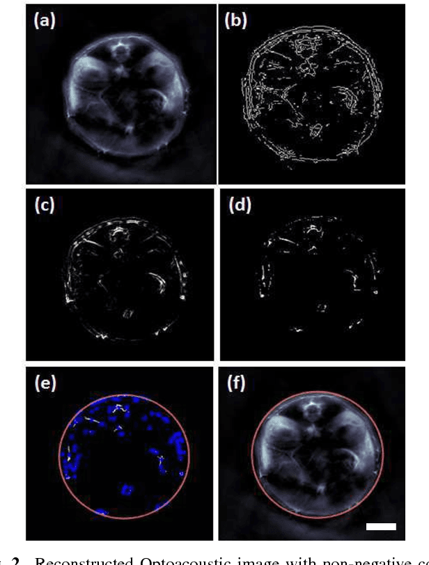
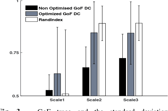
Abstract:In this article, we present a novel scheme for segmenting the image boundary (with the background) in optoacoustic small animal in vivo imaging systems. The method utilizes a multiscale edge detection algorithm to generate a binary edge map. A scale dependent morphological operation is employed to clean spurious edges. Thereafter, an ellipse is fitted to the edge map through constrained parametric transformations and iterative goodness of fit calculations. The method delimits the tissue edges through the curve fitting model, which has shown high levels of accuracy. Thus, this method enables segmentation of optoacoutic images with minimal human intervention, by eliminating need of scale selection for multiscale processing and seed point determination for contour mapping.
* Engineering in Medicine and Biology Society (EMBC), 2015 37th Annual International Conference of the IEEE (Accepted version)
 Add to Chrome
 Add to Firefox
 Add to Edge