Xosé Luís Deán-Ben
OADAT: Experimental and Synthetic Clinical Optoacoustic Data for Standardized Image Processing
Jun 17, 2022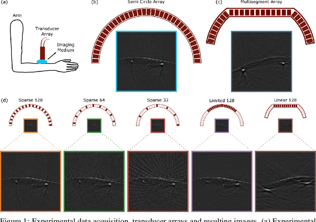

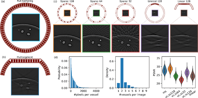
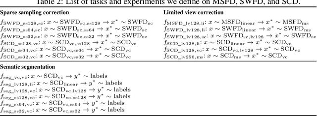
Abstract:Optoacoustic (OA) imaging is based on excitation of biological tissues with nanosecond-duration laser pulses followed by subsequent detection of ultrasound waves generated via light-absorption-mediated thermoelastic expansion. OA imaging features a powerful combination between rich optical contrast and high resolution in deep tissues. This enabled the exploration of a number of attractive new applications both in clinical and laboratory settings. However, no standardized datasets generated with different types of experimental set-up and associated processing methods are available to facilitate advances in broader applications of OA in clinical settings. This complicates an objective comparison between new and established data processing methods, often leading to qualitative results and arbitrary interpretations of the data. In this paper, we provide both experimental and synthetic OA raw signals and reconstructed image domain datasets rendered with different experimental parameters and tomographic acquisition geometries. We further provide trained neural networks to tackle three important challenges related to OA image processing, namely accurate reconstruction under limited view tomographic conditions, removal of spatial undersampling artifacts and anatomical segmentation for improved image reconstruction. Specifically, we define 18 experiments corresponding to the aforementioned challenges as benchmarks to be used as a reference for the development of more advanced processing methods.
Visual Quality Enhancement in Optoacoustic Tomography using Active Contour Segmentation Priors
Apr 11, 2016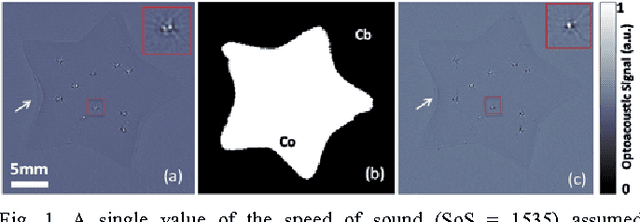
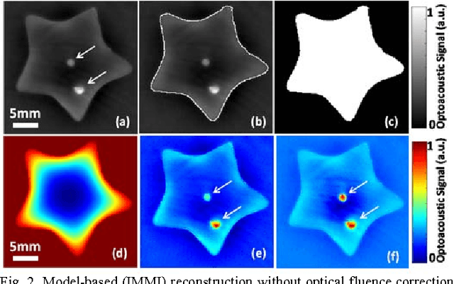
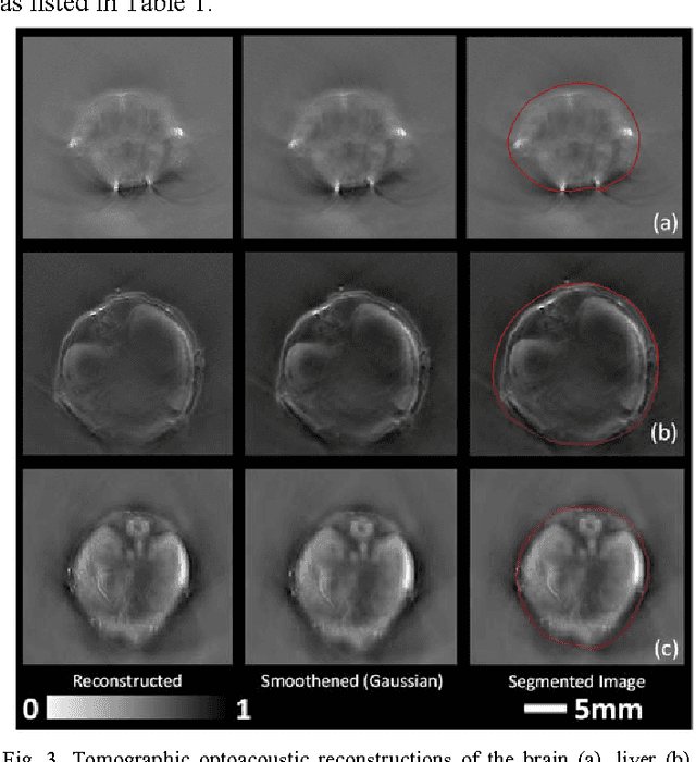
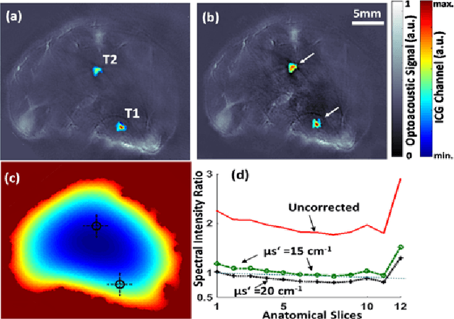
Abstract:Segmentation of biomedical images is essential for studying and characterizing anatomical structures, detection and evaluation of pathological tissues. Segmentation has been further shown to enhance the reconstruction performance in many tomographic imaging modalities by accounting for heterogeneities of the excitation field and tissue properties in the imaged region. This is particularly relevant in optoacoustic tomography, where discontinuities in the optical and acoustic tissue properties, if not properly accounted for, may result in deterioration of the imaging performance. Efficient segmentation of optoacoustic images is often hampered by the relatively low intrinsic contrast of large anatomical structures, which is further impaired by the limited angular coverage of some commonly employed tomographic imaging configurations. Herein, we analyze the performance of active contour models for boundary segmentation in cross-sectional optoacoustic tomography. The segmented mask is employed to construct a two compartment model for the acoustic and optical parameters of the imaged tissues, which is subsequently used to improve accuracy of the image reconstruction routines. The performance of the suggested segmentation and modeling approach are showcased in tissue-mimicking phantoms and small animal imaging experiments.
 Add to Chrome
 Add to Firefox
 Add to Edge