Seung Yeon Shin
Improving Segmentation and Detection of Lesions in CT Scans Using Intensity Distribution Supervision
Jul 11, 2023Abstract:We propose a method to incorporate the intensity information of a target lesion on CT scans in training segmentation and detection networks. We first build an intensity-based lesion probability (ILP) function from an intensity histogram of the target lesion. It is used to compute the probability of being the lesion for each voxel based on its intensity. Finally, the computed ILP map of each input CT scan is provided as additional supervision for network training, which aims to inform the network about possible lesion locations in terms of intensity values at no additional labeling cost. The method was applied to improve the segmentation of three different lesion types, namely, small bowel carcinoid tumor, kidney tumor, and lung nodule. The effectiveness of the proposed method on a detection task was also investigated. We observed improvements of 41.3% -> 47.8%, 74.2% -> 76.0%, and 26.4% -> 32.7% in segmenting small bowel carcinoid tumor, kidney tumor, and lung nodule, respectively, in terms of per case Dice scores. An improvement of 64.6% -> 75.5% was achieved in detecting kidney tumors in terms of average precision. The results of different usages of the ILP map and the effect of varied amount of training data are also presented.
Improving Small Lesion Segmentation in CT Scans using Intensity Distribution Supervision: Application to Small Bowel Carcinoid Tumor
Jul 29, 2022

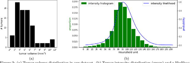
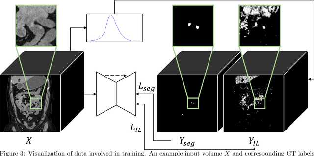
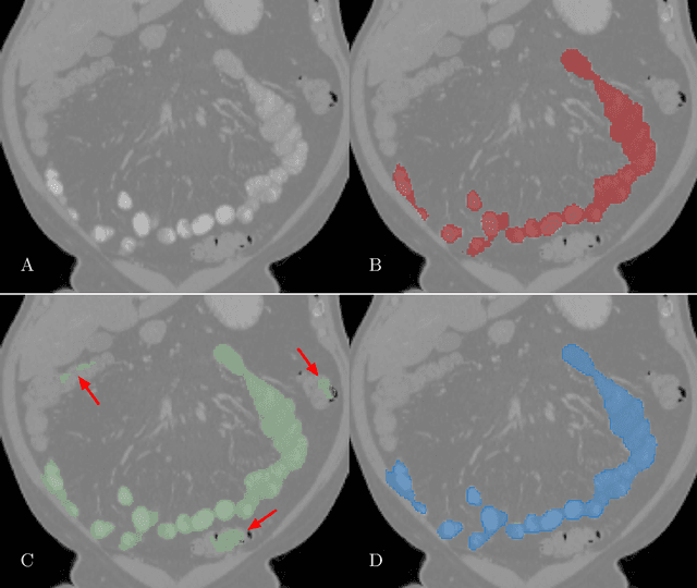
Abstract:Finding small lesions is very challenging due to lack of noticeable features, severe class imbalance, as well as the size itself. One approach to improve small lesion segmentation is to reduce the region of interest and inspect it at a higher sensitivity rather than performing it for the entire region. It is usually implemented as sequential or joint segmentation of organ and lesion, which requires additional supervision on organ segmentation. Instead, we propose to utilize an intensity distribution of a target lesion at no additional labeling cost to effectively separate regions where the lesions are possibly located from the background. It is incorporated into network training as an auxiliary task. We applied the proposed method to segmentation of small bowel carcinoid tumors in CT scans. We observed improvements for all metrics (33.5% $\rightarrow$ 38.2%, 41.3% $\rightarrow$ 47.8%, 30.0% $\rightarrow$ 35.9% for the global, per case, and per tumor Dice scores, respectively.) compared to the baseline method, which proves the validity of our idea. Our method can be one option for explicitly incorporating intensity distribution information of a target in network training.
Graph-Based Small Bowel Path Tracking with Cylindrical Constraints
Jul 29, 2022


Abstract:We present a new graph-based method for small bowel path tracking based on cylindrical constraints. A distinctive characteristic of the small bowel compared to other organs is the contact between parts of itself along its course, which makes the path tracking difficult together with the indistinct appearance of the wall. It causes the tracked path to easily cross over the walls when relying on low-level features like the wall detection. To circumvent this, a series of cylinders that are fitted along the course of the small bowel are used to guide the tracking to more reliable directions. It is implemented as soft constraints using a new cost function. The proposed method is evaluated against ground-truth paths that are all connected from start to end of the small bowel for 10 abdominal CT scans. The proposed method showed clear improvements compared to the baseline method in tracking the path without making an error. Improvements of 6.6% and 17.0%, in terms of the tracked length, were observed for two different settings related to the small bowel segmentation.
Extraction of Coronary Vessels in Fluoroscopic X-Ray Sequences Using Vessel Correspondence Optimization
Jul 28, 2022Abstract:We present a method to extract coronary vessels from fluoroscopic x-ray sequences. Given the vessel structure for the source frame, vessel correspondence candidates in the subsequent frame are generated by a novel hierarchical search scheme to overcome the aperture problem. Optimal correspondences are determined within a Markov random field optimization framework. Post-processing is performed to extract vessel branches newly visible due to the inflow of contrast agent. Quantitative and qualitative evaluation conducted on a dataset of 18 sequences demonstrates the effectiveness of the proposed method.
Deep Reinforcement Learning for Small Bowel Path Tracking using Different Types of Annotations
Jun 29, 2022

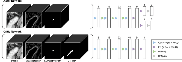

Abstract:Small bowel path tracking is a challenging problem considering its many folds and contact along its course. For the same reason, it is very costly to achieve the ground-truth (GT) path of the small bowel in 3D. In this work, we propose to train a deep reinforcement learning tracker using datasets with different types of annotations. Specifically, we utilize CT scans that have only GT small bowel segmentation as well as ones with the GT path. It is enabled by designing a unique environment that is compatible for both, including a reward definable even without the GT path. The performed experiments proved the validity of the proposed method. The proposed method holds a high degree of usability in this problem by being able to utilize the scans with weak annotations, and thus by possibly reducing the required annotation cost.
A Graph-theoretic Algorithm for Small Bowel Path Tracking in CT Scans
Oct 01, 2021

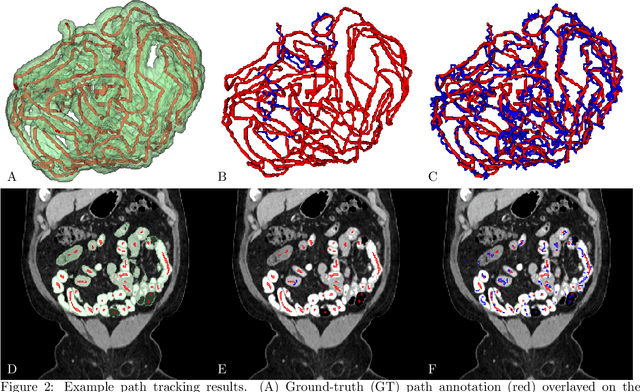
Abstract:We present a novel graph-theoretic method for small bowel path tracking. It is formulated as finding the minimum cost path between given start and end nodes on a graph that is constructed based on the bowel wall detection. We observed that a trivial solution with many short-cuts is easily made even with the wall detection, where the tracked path penetrates indistinct walls around the contact between different parts of the small bowel. Thus, we propose to include must-pass nodes in finding the path to better cover the entire course of the small bowel. The proposed method does not entail training with ground-truth paths while the previous methods do. We acquired ground-truth paths that are all connected from start to end of the small bowel for 10 abdominal CT scans, which enables the evaluation of the path tracking for the entire course of the small bowel. The proposed method showed clear improvements in terms of several metrics compared to the baseline method. The maximum length of the path that is tracked without an error per scan, by the proposed method, is above 800mm on average.
Unsupervised Domain Adaptation for Small Bowel Segmentation using Disentangled Representation
Jul 06, 2021Abstract:We present a novel unsupervised domain adaptation method for small bowel segmentation based on feature disentanglement. To make the domain adaptation more controllable, we disentangle intensity and non-intensity features within a unique two-stream auto-encoding architecture, and selectively adapt the non-intensity features that are believed to be more transferable across domains. The segmentation prediction is performed by aggregating the disentangled features. We evaluated our method using intravenous contrast-enhanced abdominal CT scans with and without oral contrast, which are used as source and target domains, respectively. The proposed method showed clear improvements in terms of three different metrics compared to other domain adaptation methods that are without the feature disentanglement. The method brings small bowel segmentation closer to clinical application.
Deep Small Bowel Segmentation with Cylindrical Topological Constraints
Jul 16, 2020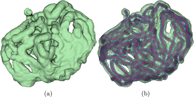
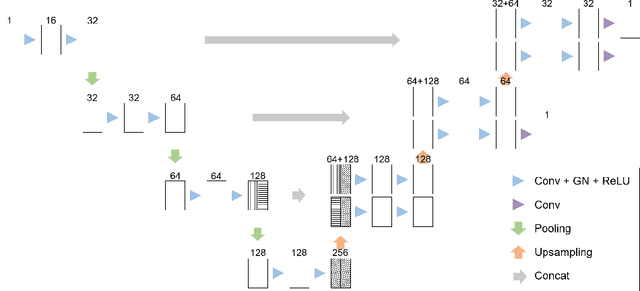

Abstract:We present a novel method for small bowel segmentation where a cylindrical topological constraint based on persistent homology is applied. To address the touching issue which could break the applied constraint, we propose to augment a network with an additional branch to predict an inner cylinder of the small bowel. Since the inner cylinder is free of the touching issue, a cylindrical shape constraint applied on this augmented branch guides the network to generate a topologically correct segmentation. For strict evaluation, we achieved an abdominal computed tomography dataset with dense segmentation ground-truths. The proposed method showed clear improvements in terms of four different metrics compared to the baseline method, and also showed the statistical significance from a paired t-test.
Deep Vessel Segmentation By Learning Graphical Connectivity
Jun 06, 2018
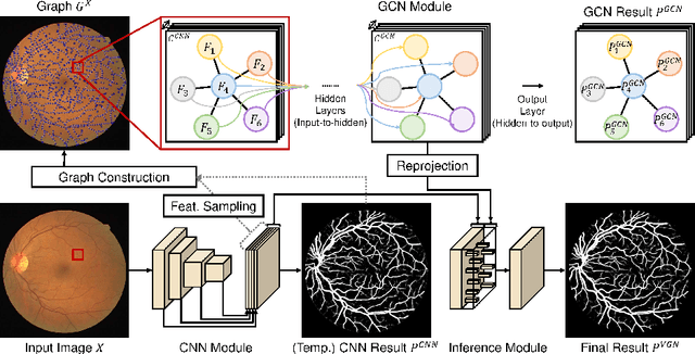
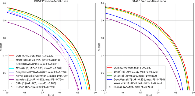
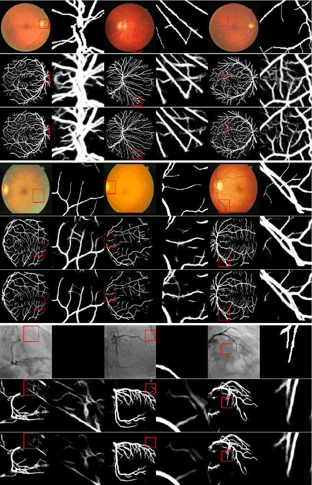
Abstract:We propose a novel deep-learning-based system for vessel segmentation. Existing methods using CNNs have mostly relied on local appearances learned on the regular image grid, without considering the graphical structure of vessel shape. To address this, we incorporate a graph convolutional network into a unified CNN architecture, where the final segmentation is inferred by combining the different types of features. The proposed method can be applied to expand any type of CNN-based vessel segmentation method to enhance the performance. Experiments show that the proposed method outperforms the current state-of-the-art methods on two retinal image datasets as well as a coronary artery X-ray angiography dataset.
Joint Weakly and Semi-Supervised Deep Learning for Localization and Classification of Masses in Breast Ultrasound Images
Oct 10, 2017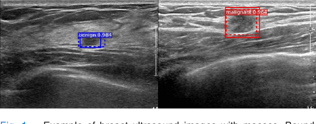
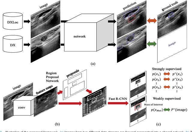
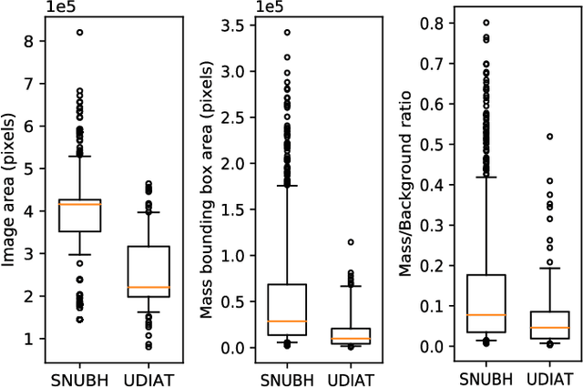

Abstract:We propose a framework for localization and classification of masses in breast ultrasound (BUS) images. In particular, we simultaneously use a weakly annotated dataset and a relatively small strongly annotated dataset to train a convolutional neural network detector. We have experimentally found that mass detectors trained with small, strongly annotated datasets are easily overfitted, whereas those trained with large, weakly annotated datasets present a non-trivial problem. To overcome these problems, we jointly use datasets with different characteristics in a hybrid manner. Consequently, a sophisticated weakly and semi-supervised training scenario is introduced with appropriate training loss selection. Experimental results show that the proposed method successfully localizes and classifies masses while requiring less effort in annotation work. The influences of each component in the proposed framework are also validated by conducting an ablative analysis. Although the proposed method is intended for masses in BUS images, it can also be applied as a general framework to train computer-aided detection and diagnosis systems for a wide variety of image modalities, target organs, and diseases.
 Add to Chrome
 Add to Firefox
 Add to Edge