Samira Vafay Eslahi
Dual Prompting for Diverse Count-level PET Denoising
May 05, 2025Abstract:The to-be-denoised positron emission tomography (PET) volumes are inherent with diverse count levels, which imposes challenges for a unified model to tackle varied cases. In this work, we resort to the recently flourished prompt learning to achieve generalizable PET denoising with different count levels. Specifically, we propose dual prompts to guide the PET denoising in a divide-and-conquer manner, i.e., an explicitly count-level prompt to provide the specific prior information and an implicitly general denoising prompt to encode the essential PET denoising knowledge. Then, a novel prompt fusion module is developed to unify the heterogeneous prompts, followed by a prompt-feature interaction module to inject prompts into the features. The prompts are able to dynamically guide the noise-conditioned denoising process. Therefore, we are able to efficiently train a unified denoising model for various count levels, and deploy it to different cases with personalized prompts. We evaluated on 1940 low-count PET 3D volumes with uniformly randomly selected 13-22\% fractions of events from 97 $^{18}$F-MK6240 tau PET studies. It shows our dual prompting can largely improve the performance with informed count-level and outperform the count-conditional model.
Free-breathing 3D cardiac extracellular volume (ECV) mapping using a linear tangent space alignment (LTSA) model
Aug 22, 2024


Abstract:$\textbf{Purpose:}$ To develop a new method for free-breathing 3D extracellular volume (ECV) mapping of the whole heart at 3T. $\textbf{Methods:}$ A free-breathing 3D cardiac ECV mapping method was developed at 3T. T1 mapping was performed before and after contrast agent injection using a free-breathing ECG-gated inversion-recovery sequence with spoiled gradient echo readout. A linear tangent space alignment (LTSA) model-based method was used to reconstruct high-frame-rate dynamic images from (k,t)-space data sparsely sampled along a random stack-of-stars trajectory. Joint T1 and transmit B1 estimation was performed voxel-by-voxel for pre- and post-contrast T1 mapping. To account for the time-varying T1 after contrast agent injection, a linearly time-varying T1 model was introduced for post-contrast T1 mapping. ECV maps were generated by aligning pre- and post-contrast T1 maps through affine transformation. $\textbf{Results:}$ The feasibility of the proposed method was demonstrated using in vivo studies with six healthy volunteers at 3T. We obtained 3D ECV maps at a spatial resolution of 1.9$\times$1.9$\times$4.5 $mm^{3}$ and a FOV of 308$\times$308$\times$144 $mm^{3}$, with a scan time of 10.1$\pm$1.4 and 10.6$\pm$1.6 min before and after contrast agent injection, respectively. The ECV maps and the pre- and post-contrast T1 maps obtained by the proposed method were in good agreement with the 2D MOLLI method both qualitatively and quantitatively. $\textbf{Conclusion:}$ The proposed method allows for free-breathing 3D ECV mapping of the whole heart within a practically feasible imaging time. The estimated ECV values from the proposed method were comparable to those from the existing method. $\textbf{Keywords:}$ cardiac extracellular volume (ECV) mapping, cardiac T1 mapping, linear tangent space alignment (LTSA), manifold learning
ERNAS: An Evolutionary Neural Architecture Search for Magnetic Resonance Image Reconstructions
Jun 15, 2022


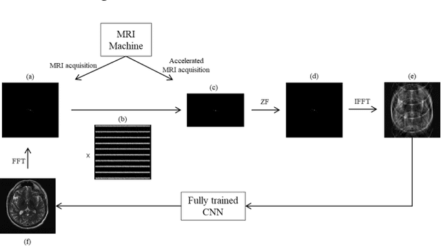
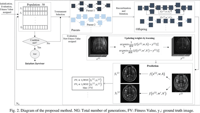
Abstract:Magnetic resonance imaging (MRI) is one of the noninvasive imaging modalities that can produce high-quality images. However, the scan procedure is relatively slow, which causes patient discomfort and motion artifacts in images. Accelerating MRI hardware is constrained by physical and physiological limitations. A popular alternative approach to accelerated MRI is to undersample the k-space data. While undersampling speeds up the scan procedure, it generates artifacts in the images, and advanced reconstruction algorithms are needed to produce artifact-free images. Recently deep learning has emerged as a promising MRI reconstruction method to address this problem. However, straightforward adoption of the existing deep learning neural network architectures in MRI reconstructions is not usually optimal in terms of efficiency and reconstruction quality. In this work, MRI reconstruction from undersampled data was carried out using an optimized neural network using a novel evolutionary neural architecture search algorithm. Brain and knee MRI datasets show that the proposed algorithm outperforms manually designed neural network-based MR reconstruction models.
 Add to Chrome
 Add to Firefox
 Add to Edge