Reed Chen
Emulating Clinical Quality Muscle B-mode Ultrasound Images from Plane Wave Images Using a Two-Stage Machine Learning Model
Dec 07, 2024Abstract:Research ultrasound scanners such as the Verasonics Vantage often lack the advanced image processing algorithms used by clinical systems. Image quality is even lower in plane wave imaging - often used for shear wave elasticity imaging (SWEI) - which sacrifices spatial resolution for temporal resolution. As a result, delay-and-summed images acquired from SWEI have limited interpretability. In this project, a two-stage machine learning model was trained to enhance single plane wave images of muscle acquired with a Verasonics Vantage system. The first stage of the model consists of a U-Net trained to emulate plane wave compounding, histogram matching, and unsharp masking using paired images. The second stage consists of a CycleGAN trained to emulate clinical muscle B-modes using unpaired images. This two-stage model was implemented on the Verasonics Vantage research ultrasound scanner, and its ability to provide high-speed image formation at a frame rate of 28.5 +/- 0.6 FPS from a single plane wave transmit was demonstrated. A reader study with two physicians demonstrated that these processed images had significantly greater structural fidelity and less speckle than the original plane wave images.
Cryo-ZSSR: multiple-image super-resolution based on deep internal learning
Nov 22, 2020


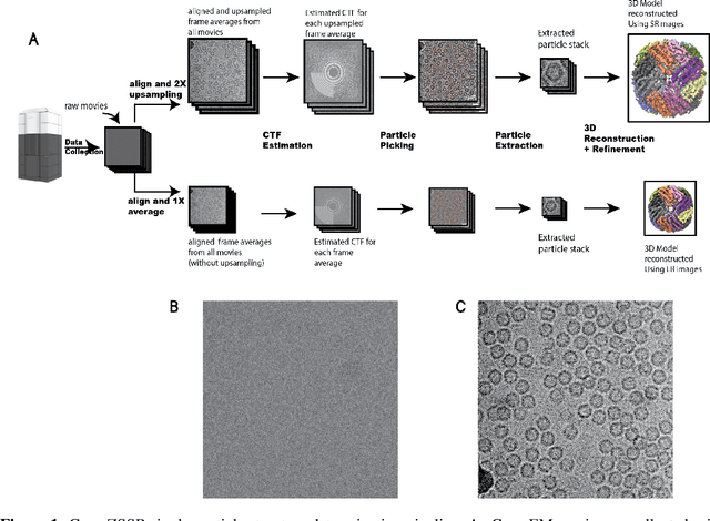
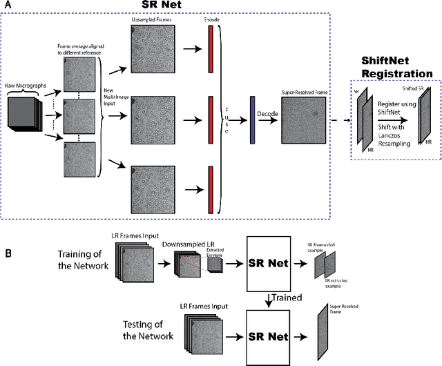
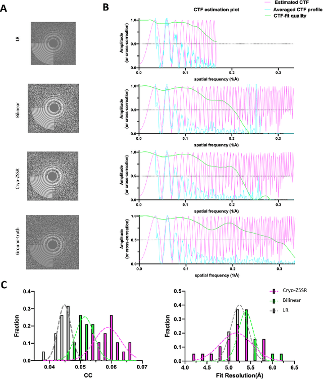
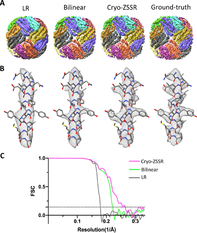
Abstract:Single-particle cryo-electron microscopy (cryo-EM) is an emerging imaging modality capable of visualizing proteins and macro-molecular complexes at near-atomic resolution. The low electron-doses used to prevent sample radiation damage, result in images where the power of the noise is 100 times greater than the power of the signal. To overcome the low-SNRs, hundreds of thousands of particle projections acquired over several days of data collection are averaged in 3D to determine the structure of interest. Meanwhile, recent image super-resolution (SR) techniques based on neural networks have shown state of the art performance on natural images. Building on these advances, we present a multiple-image SR algorithm based on deep internal learning designed specifically to work under low-SNR conditions. Our approach leverages the internal image statistics of cryo-EM movies and does not require training on ground-truth data. When applied to a single-particle dataset of apoferritin, we show that the resolution of 3D structures obtained from SR micrographs can surpass the limits imposed by the imaging system. Our results indicate that the combination of low magnification imaging with image SR has the potential to accelerate cryo-EM data collection without sacrificing resolution.
 Add to Chrome
 Add to Firefox
 Add to Edge