R. V. Paul Chan
A portable widefield fundus camera with high dynamic range imaging capability
Dec 20, 2022Abstract:Fundus photography is indispensable for clinical detection and management of eye diseases. Limited image contrast and field of view (FOV) are common limitations of conventional fundus cameras, making it difficult to detect subtle abnormalities at the early stages of eye diseases. Further improvements of image contrast and FOV coverage are important to improve early disease detection and reliable treatment assessment. We report here a portable fundus camera, with a wide FOV and high dynamic range (HDR) imaging capabilities. Miniaturized indirect ophthalmoscopy illumination was employed to achieve the portable design for nonmydriatic, widefield fundus photography. Orthogonal polarization control was used to eliminate illumination reflectance artifact. With independent power controls, three fundus images were sequentially acquired and fused to achieve HDR function for local image contrast enhancement. A 101{\deg} eye-angle (67{\deg} visual-angle) snapshot FOV was achieved for nonmydriatic fundus photography. The effective FOV can be readily expanded up to 190{\deg} eye-angle (134{\deg} visual-angle) with the aid of a fixation target, without the need of pharmacologic pupillary dilation. The effectiveness of HDR imaging was validated with both normal healthy and pathologic eyes, compared to a conventional fundus camera.
Not Color Blind: AI Predicts Racial Identity from Black and White Retinal Vessel Segmentations
Sep 28, 2021


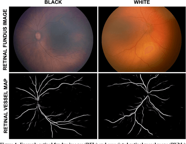
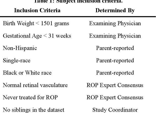
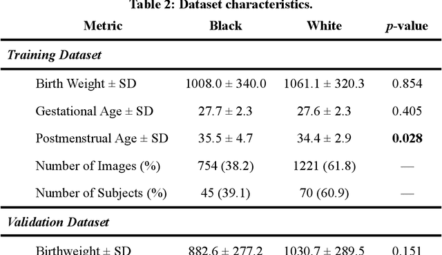
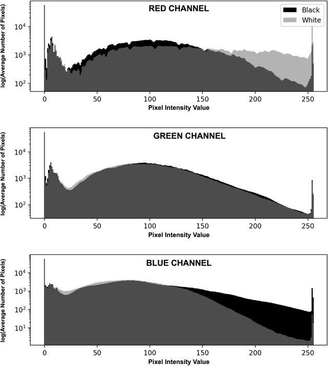
Abstract:Background: Artificial intelligence (AI) may demonstrate racial bias when skin or choroidal pigmentation is present in medical images. Recent studies have shown that convolutional neural networks (CNNs) can predict race from images that were not previously thought to contain race-specific features. We evaluate whether grayscale retinal vessel maps (RVMs) of patients screened for retinopathy of prematurity (ROP) contain race-specific features. Methods: 4095 retinal fundus images (RFIs) were collected from 245 Black and White infants. A U-Net generated RVMs from RFIs, which were subsequently thresholded, binarized, or skeletonized. To determine whether RVM differences between Black and White eyes were physiological, CNNs were trained to predict race from color RFIs, raw RVMs, and thresholded, binarized, or skeletonized RVMs. Area under the precision-recall curve (AUC-PR) was evaluated. Findings: CNNs predicted race from RFIs near perfectly (image-level AUC-PR: 0.999, subject-level AUC-PR: 1.000). Raw RVMs were almost as informative as color RFIs (image-level AUC-PR: 0.938, subject-level AUC-PR: 0.995). Ultimately, CNNs were able to detect whether RFIs or RVMs were from Black or White babies, regardless of whether images contained color, vessel segmentation brightness differences were nullified, or vessel segmentation widths were normalized. Interpretation: AI can detect race from grayscale RVMs that were not thought to contain racial information. Two potential explanations for these findings are that: retinal vessels physiologically differ between Black and White babies or the U-Net segments the retinal vasculature differently for various fundus pigmentations. Either way, the implications remain the same: AI algorithms have potential to demonstrate racial bias in practice, even when preliminary attempts to remove such information from the underlying images appear to be successful.
 Add to Chrome
 Add to Firefox
 Add to Edge