Philipp Ernst
Primal-Dual UNet for Sparse View Cone Beam Computed Tomography Volume Reconstruction
May 11, 2022
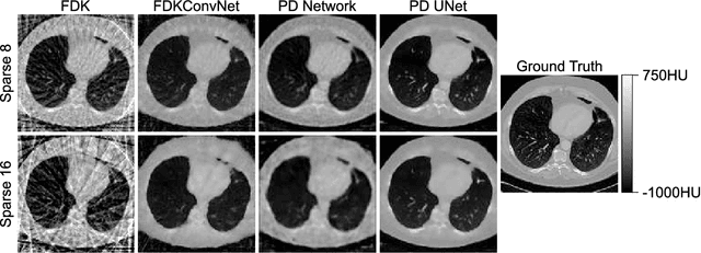
Abstract:In this paper, the Primal-Dual UNet for sparse view CT reconstruction is modified to be applicable to cone beam projections and perform reconstructions of entire volumes instead of slices. Experiments show that the PSNR of the proposed method is increased by 10dB compared to the direct FDK reconstruction and almost 3dB compared to the modified original Primal-Dual Network when using only 23 projections. The presented network is not optimized wrt. memory consumption or hyperparameters but merely serves as a proof of concept and is limited to low resolution projections and volumes.
Dual Branch Prior-SegNet: CNN for Interventional CBCT using Planning Scan and Auxiliary Segmentation Loss
May 11, 2022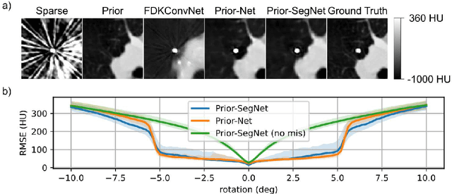
Abstract:This paper proposes an extension to the Dual Branch Prior-Net for sparse view interventional CBCT reconstruction incorporating a high quality planning scan. An additional head learns to segment interventional instruments and thus guides the reconstruction task. The prior scans are misaligned by up to +-5deg in-plane during training. Experiments show that the proposed model, Dual Branch Prior-SegNet, significantly outperforms any other evaluated model by >2.8dB PSNR. It also stays robust wrt. rotations of up to +-5.5deg.
Sinogram upsampling using Primal-Dual UNet for undersampled CT and radial MRI reconstruction
Dec 26, 2021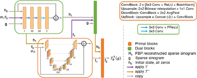

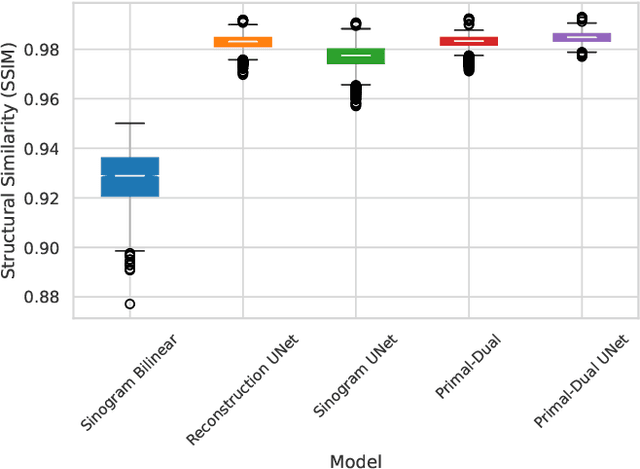
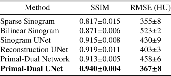
Abstract:CT and MRI are two widely used clinical imaging modalities for non-invasive diagnosis. However, both of these modalities come with certain problems. CT uses harmful ionising radiation, and MRI suffers from slow acquisition speed. Both problems can be tackled by undersampling, such as sparse sampling. However, such undersampled data leads to lower resolution and introduces artefacts. Several techniques, including deep learning based methods, have been proposed to reconstruct such data. However, the undersampled reconstruction problem for these two modalities was always considered as two different problems and tackled separately by different research works. This paper proposes a unified solution for both sparse CT and undersampled radial MRI reconstruction, achieved by applying Fourier transform-based pre-processing on the radial MRI and then reconstructing both modalities using sinogram upsampling combined with filtered back-projection. The Primal-Dual network is a deep learning based method for reconstructing sparsely-sampled CT data. This paper introduces Primal-Dual UNet, which improves the Primal-Dual network in terms of accuracy and reconstruction speed. The proposed method resulted in an average SSIM of 0.932 while performing sparse CT reconstruction for fan-beam geometry with a sparsity level of 16, achieving a statistically significant improvement over the previous model, which resulted in 0.919. Furthermore, the proposed model resulted in 0.903 and 0.957 average SSIM while reconstructing undersampled brain and abdominal MRI data with an acceleration factor of 16 - statistically significant improvements over the original model, which resulted in 0.867 and 0.949. Finally, this paper shows that the proposed network not only improves the overall image quality, but also improves the image quality for the regions-of-interest; as well as generalises better in presence of a needle.
Dual Skip Connections Minimize the False Positive Rate of Lung Nodule Detection in CT images
Oct 25, 2021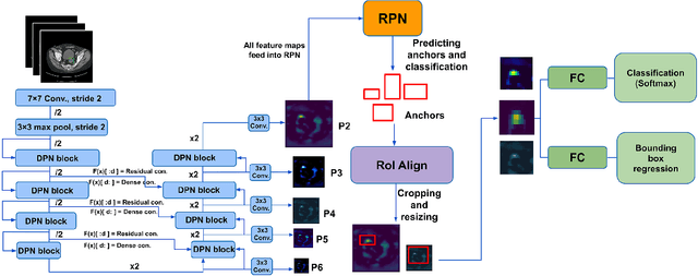
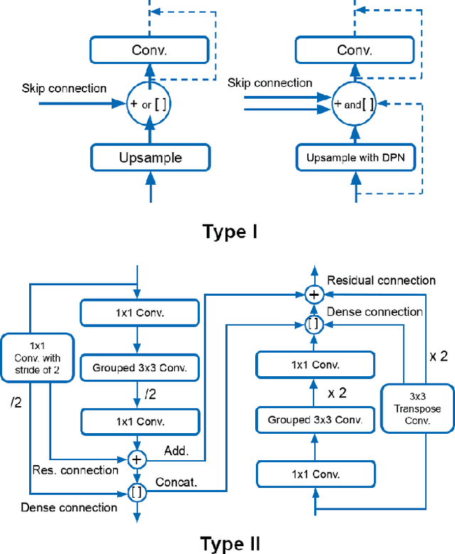
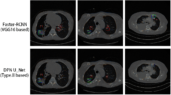

Abstract:Pulmonary cancer is one of the most commonly diagnosed and fatal cancers and is often diagnosed by incidental findings on computed tomography. Automated pulmonary nodule detection is an essential part of computer-aided diagnosis, which is still facing great challenges and difficulties to quickly and accurately locate the exact nodules' positions. This paper proposes a dual skip connection upsampling strategy based on Dual Path network in a U-Net structure generating multiscale feature maps, which aims to minimize the ratio of false positives and maximize the sensitivity for lesion detection of nodules. The results show that our new upsampling strategy improves the performance by having 85.3% sensitivity at 4 FROC per image compared to 84.2% for the regular upsampling strategy or 81.2% for VGG16-based Faster-R-CNN.
CHAOS Challenge -- Combined (CT-MR) Healthy Abdominal Organ Segmentation
Jan 17, 2020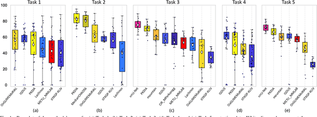
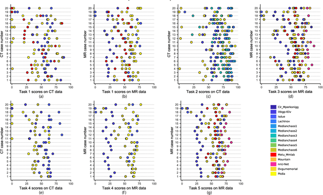

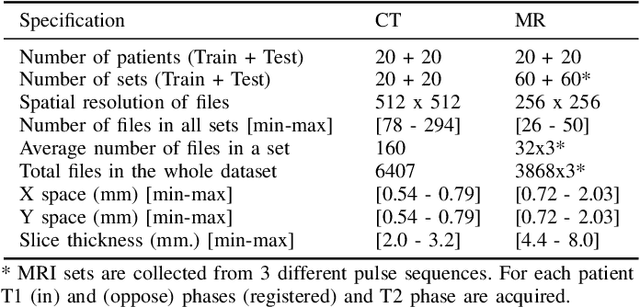
Abstract:Segmentation of abdominal organs has been a comprehensive, yet unresolved, research field for many years. In the last decade, intensive developments in deep learning (DL) have introduced new state-of-the-art segmentation systems. Despite outperforming the overall accuracy of existing systems, the effects of DL model properties and parameters on the performance is hard to interpret. This makes comparative analysis a necessary tool to achieve explainable studies and systems. Moreover, the performance of DL for emerging learning approaches such as cross-modality and multi-modal tasks have been rarely discussed. In order to expand the knowledge in these topics, CHAOS -- Combined (CT-MR) Healthy Abdominal Organ Segmentation challenge has been organized in the IEEE International Symposium on Biomedical Imaging (ISBI), 2019, in Venice, Italy. Despite a large number of the previous abdomen related challenges, the majority of which are focused on tumor/lesion detection and/or classification with a single modality, CHAOS provides both abdominal CT and MR data from healthy subjects. Five different and complementary tasks have been designed to analyze the capabilities of the current approaches from multiple perspectives. The results are investigated thoroughly, compared with manual annotations and interactive methods. The outcomes are reported in detail to reflect the latest advancements in the field. CHAOS challenge and data will be available online to provide a continuous benchmark resource for segmentation.
 Add to Chrome
 Add to Firefox
 Add to Edge