Muhammad Arsalan
Feature Enhancer Segmentation Network (FES-Net) for Vessel Segmentation
Sep 07, 2023


Abstract:Diseases such as diabetic retinopathy and age-related macular degeneration pose a significant risk to vision, highlighting the importance of precise segmentation of retinal vessels for the tracking and diagnosis of progression. However, existing vessel segmentation methods that heavily rely on encoder-decoder structures struggle to capture contextual information about retinal vessel configurations, leading to challenges in reconciling semantic disparities between encoder and decoder features. To address this, we propose a novel feature enhancement segmentation network (FES-Net) that achieves accurate pixel-wise segmentation without requiring additional image enhancement steps. FES-Net directly processes the input image and utilizes four prompt convolutional blocks (PCBs) during downsampling, complemented by a shallow upsampling approach to generate a binary mask for each class. We evaluate the performance of FES-Net on four publicly available state-of-the-art datasets: DRIVE, STARE, CHASE, and HRF. The evaluation results clearly demonstrate the superior performance of FES-Net compared to other competitive approaches documented in the existing literature.
MKIS-Net: A Light-Weight Multi-Kernel Network for Medical Image Segmentation
Oct 15, 2022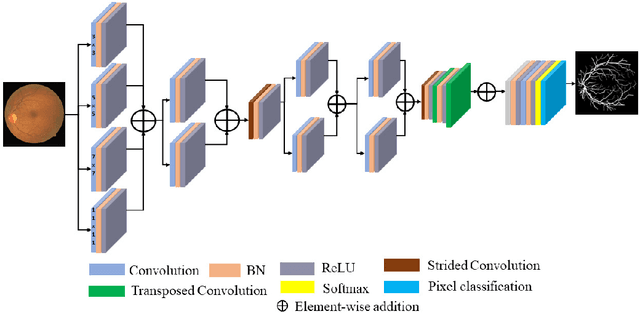
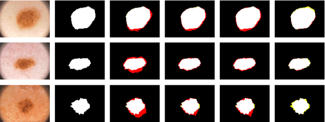
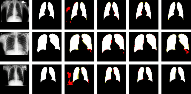
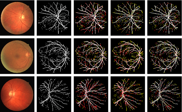
Abstract:Image segmentation is an important task in medical imaging. It constitutes the backbone of a wide variety of clinical diagnostic methods, treatments, and computer-aided surgeries. In this paper, we propose a multi-kernel image segmentation net (MKIS-Net), which uses multiple kernels to create an efficient receptive field and enhance segmentation performance. As a result of its multi-kernel design, MKIS-Net is a light-weight architecture with a small number of trainable parameters. Moreover, these multi-kernel receptive fields also contribute to better segmentation results. We demonstrate the efficacy of MKIS-Net on several tasks including segmentation of retinal vessels, skin lesion segmentation, and chest X-ray segmentation. The performance of the proposed network is quite competitive, and often superior, in comparison to state-of-the-art methods. Moreover, in some cases MKIS-Net has more than an order of magnitude fewer trainable parameters than existing medical image segmentation alternatives and is at least four times smaller than other light-weight architectures.
A Residual Encoder-Decoder Network for Segmentation of Retinal Image-Based Exudates in Diabetic Retinopathy Screening
Jan 16, 2022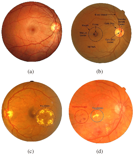
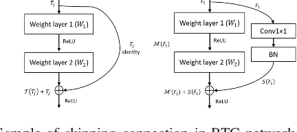
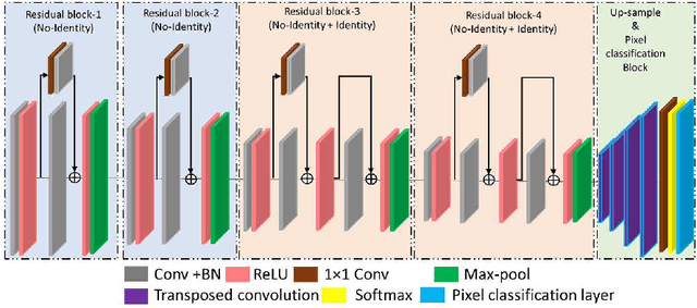

Abstract:Diabetic retinopathy refers to the pathology of the retina induced by diabetes and is one of the leading causes of preventable blindness in the world. Early detection of diabetic retinopathy is critical to avoid vision problem through continuous screening and treatment. In traditional clinical practice, the involved lesions are manually detected using photographs of the fundus. However, this task is cumbersome and time-consuming and requires intense effort due to the small size of lesion and low contrast of the images. Thus, computer-assisted diagnosis of diabetic retinopathy based on the detection of red lesions is actively being explored recently. In this paper, we present a convolutional neural network with residual skip connection for the segmentation of exudates in retinal images. To improve the performance of network architecture, a suitable image augmentation technique is used. The proposed network can robustly segment exudates with high accuracy, which makes it suitable for diabetic retinopathy screening. Comparative performance analysis of three benchmark databases: HEI-MED, E-ophtha, and DiaretDB1 is presented. It is shown that the proposed method achieves accuracy (0.98, 0.99, 0.98) and sensitivity (0.97, 0.92, and 0.95) on E-ophtha, HEI-MED, and DiaReTDB1, respectively.
 Add to Chrome
 Add to Firefox
 Add to Edge