Mina Khoshdeli
Efficient and generalizable prediction of molecular alterations in multiple cancer cohorts using H&E whole slide images
Jul 22, 2024Abstract:Molecular testing of tumor samples for targetable biomarkers is restricted by a lack of standardization, turnaround-time, cost, and tissue availability across cancer types. Additionally, targetable alterations of low prevalence may not be tested in routine workflows. Algorithms that predict DNA alterations from routinely generated hematoxylin and eosin (H&E)-stained images could prioritize samples for confirmatory molecular testing. Costs and the necessity of a large number of samples containing mutations limit approaches that train individual algorithms for each alteration. In this work, models were trained for simultaneous prediction of multiple DNA alterations from H&E images using a multi-task approach. Compared to biomarker-specific models, this approach performed better on average, with pronounced gains for rare mutations. The models reasonably generalized to independent temporal-holdout, externally-stained, and multi-site TCGA test sets. Additionally, whole slide image embeddings derived using multi-task models demonstrated strong performance in downstream tasks that were not a part of training. Overall, this is a promising approach to develop clinically useful algorithms that provide multiple actionable predictions from a single slide.
Deep Learning Models Delineates Multiple Nuclear Phenotypes in H&E Stained Histology Sections
Feb 14, 2018


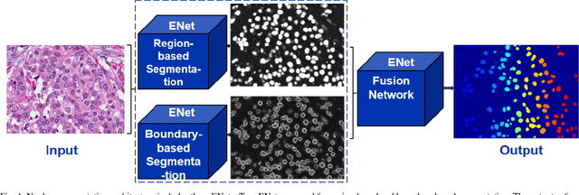
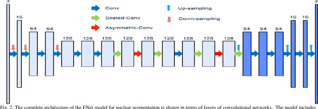
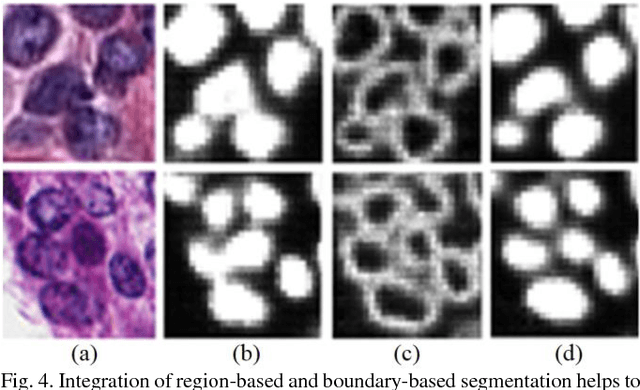
Abstract:Nuclear segmentation is an important step for profiling aberrant regions of histology sections. However, segmentation is a complex problem as a result of variations in nuclear geometry (e.g., size, shape), nuclear type (e.g., epithelial, fibroblast), and nuclear phenotypes (e.g., vesicular, aneuploidy). The problem is further complicated as a result of variations in sample preparation. It is shown and validated that fusion of very deep convolutional networks overcomes (i) complexities associated with multiple nuclear phenotypes, and (ii) separation of overlapping nuclei. The fusion relies on integrating of networks that learn region- and boundary-based representations. The system has been validated on a diverse set of nuclear phenotypes that correspond to the breast and brain histology sections.
 Add to Chrome
 Add to Firefox
 Add to Edge