Maxine Tan
A new Time-decay Radiomics Integrated Network (TRINet) for short-term breast cancer risk prediction
Dec 04, 2024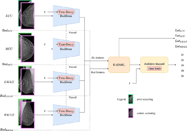
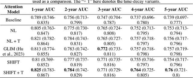
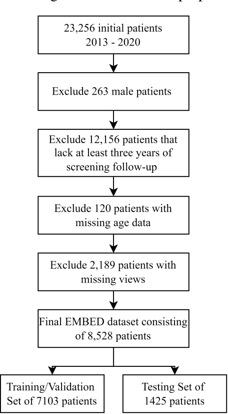
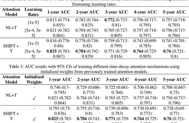
Abstract:To facilitate early detection of breast cancer, there is a need to develop short-term risk prediction schemes that can prescribe personalized/individualized screening mammography regimens for women. In this study, we propose a new deep learning architecture called TRINet that implements time-decay attention to focus on recent mammographic screenings, as current models do not account for the relevance of newer images. We integrate radiomic features with an Attention-based Multiple Instance Learning (AMIL) framework to weigh and combine multiple views for better risk estimation. In addition, we introduce a continual learning approach with a new label assignment strategy based on bilateral asymmetry to make the model more adaptable to asymmetrical cancer indicators. Finally, we add a time-embedded additive hazard layer to perform dynamic, multi-year risk forecasting based on individualized screening intervals. We used two public datasets, namely 8,528 patients from the American EMBED dataset and 8,723 patients from the Swedish CSAW dataset in our experiments. Evaluation results on the EMBED test set show that our approach significantly outperforms state-of-the-art models, achieving AUC scores of 0.851, 0.811, 0.796, 0.793, and 0.789 across 1-, 2-, to 5-year intervals, respectively. Our results underscore the importance of integrating temporal attention, radiomic features, time embeddings, bilateral asymmetry, and continual learning strategies, providing a more adaptive and precise tool for short-term breast cancer risk prediction.
RADIFUSION: A multi-radiomics deep learning based breast cancer risk prediction model using sequential mammographic images with image attention and bilateral asymmetry refinement
Apr 01, 2023


Abstract:Breast cancer is a significant public health concern and early detection is critical for triaging high risk patients. Sequential screening mammograms can provide important spatiotemporal information about changes in breast tissue over time. In this study, we propose a deep learning architecture called RADIFUSION that utilizes sequential mammograms and incorporates a linear image attention mechanism, radiomic features, a new gating mechanism to combine different mammographic views, and bilateral asymmetry-based finetuning for breast cancer risk assessment. We evaluate our model on a screening dataset called Cohort of Screen-Aged Women (CSAW) dataset. Based on results obtained on the independent testing set consisting of 1,749 women, our approach achieved superior performance compared to other state-of-the-art models with area under the receiver operating characteristic curves (AUCs) of 0.905, 0.872 and 0.866 in the three respective metrics of 1-year AUC, 2-year AUC and > 2-year AUC. Our study highlights the importance of incorporating various deep learning mechanisms, such as image attention, radiomic features, gating mechanism, and bilateral asymmetry-based fine-tuning, to improve the accuracy of breast cancer risk assessment. We also demonstrate that our model's performance was enhanced by leveraging spatiotemporal information from sequential mammograms. Our findings suggest that RADIFUSION can provide clinicians with a powerful tool for breast cancer risk assessment.
CASPIANET++: A Multidimensional Channel-Spatial Asymmetric Attention Network with Noisy Student Curriculum Learning Paradigm for Brain Tumor Segmentation
Jul 08, 2021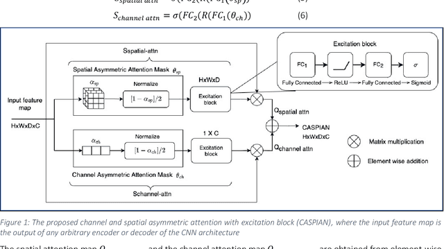
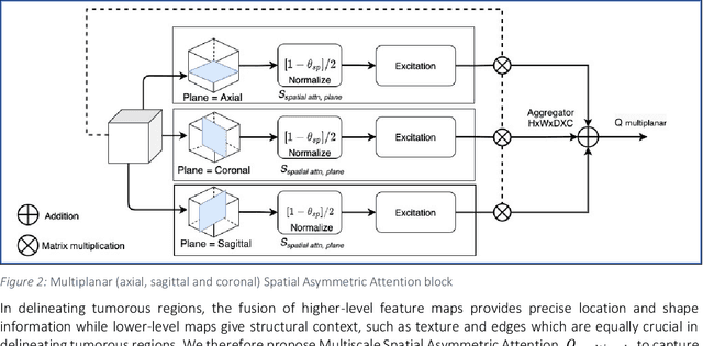
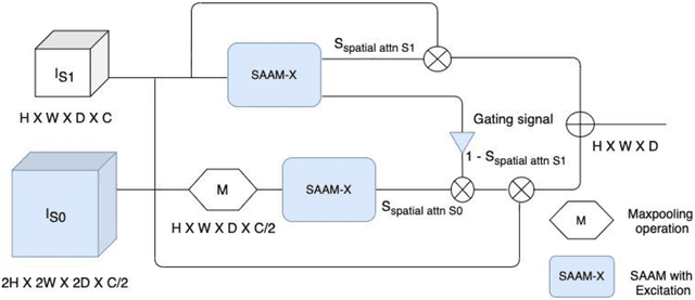
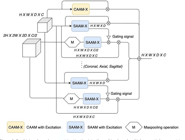
Abstract:Convolutional neural networks (CNNs) have been used quite successfully for semantic segmentation of brain tumors. However, current CNNs and attention mechanisms are stochastic in nature and neglect the morphological indicators used by radiologists to manually annotate regions of interest. In this paper, we introduce a channel and spatial wise asymmetric attention (CASPIAN) by leveraging the inherent structure of tumors to detect regions of saliency. To demonstrate the efficacy of our proposed layer, we integrate this into a well-established convolutional neural network (CNN) architecture to achieve higher Dice scores, with less GPU resources. Also, we investigate the inclusion of auxiliary multiscale and multiplanar attention branches to increase the spatial context crucial in semantic segmentation tasks. The resulting architecture is the new CASPIANET++, which achieves Dice Scores of 91.19% whole tumor, 87.6% for tumor core and 81.03% for enhancing tumor. Furthermore, driven by the scarcity of brain tumor data, we investigate the Noisy Student method for segmentation tasks. Our new Noisy Student Curriculum Learning paradigm, which infuses noise incrementally to increase the complexity of the training images exposed to the network, further boosts the enhancing tumor region to 81.53%. Additional validation performed on the BraTS2020 data shows that the Noisy Student Curriculum Learning method works well without any additional training or finetuning.
3D Axial-Attention for Lung Nodule Classification
Jan 02, 2021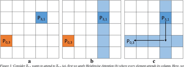

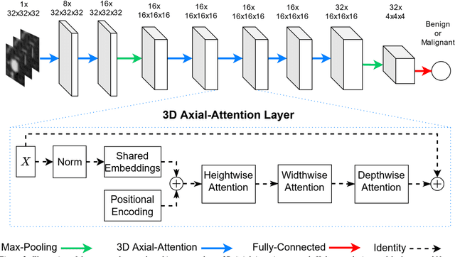
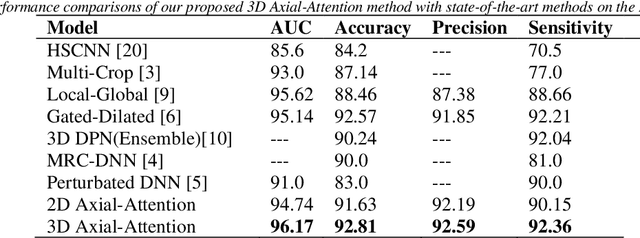
Abstract:Purpose: In recent years, Non-Local based methods have been successfully applied to lung nodule classification. However, these methods offer 2D attention or a limited 3D attention to low-resolution feature maps. Moreover, they still depend on a convenient local filter such as convolution as full 3D attention is expensive to compute and requires a big dataset, which might not be available. Methods: We propose to use 3D Axial-Attention, which requires a fraction of the computing power of a regular Non-Local network. Additionally, we solve the position invariant problem of the Non-Local network by proposing adding 3D positional encoding to shared embeddings. Results: We validated the proposed method on the LIDC-IDRI dataset by following a rigorous experimental setup using only nodules annotated by at least three radiologists. Our results show that the 3D Axial-Attention model achieves state-of-the-art performance on all evaluation metrics including AUC and Accuracy. Conclusions: The proposed model provides full 3D attention effectively, which can be used in all layers without the need for local filters. The experimental results show the importance of full 3D attention for classifying lung nodules.
A new semi-supervised self-training method for lung cancer prediction
Dec 17, 2020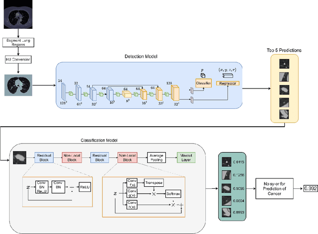


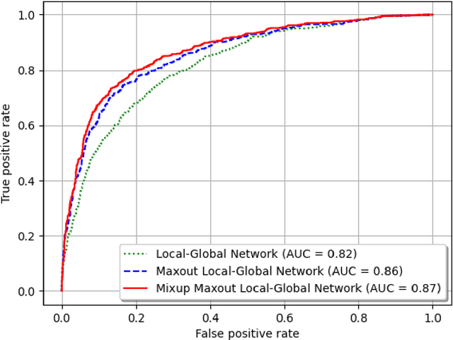
Abstract:Background and Objective: Early detection of lung cancer is crucial as it has high mortality rate with patients commonly present with the disease at stage 3 and above. There are only relatively few methods that simultaneously detect and classify nodules from computed tomography (CT) scans. Furthermore, very few studies have used semi-supervised learning for lung cancer prediction. This study presents a complete end-to-end scheme to detect and classify lung nodules using the state-of-the-art Self-training with Noisy Student method on a comprehensive CT lung screening dataset of around 4,000 CT scans. Methods: We used three datasets, namely LUNA16, LIDC and NLST, for this study. We first utilise a three-dimensional deep convolutional neural network model to detect lung nodules in the detection stage. The classification model known as Maxout Local-Global Network uses non-local networks to detect global features including shape features, residual blocks to detect local features including nodule texture, and a Maxout layer to detect nodule variations. We trained the first Self-training with Noisy Student model to predict lung cancer on the unlabelled NLST datasets. Then, we performed Mixup regularization to enhance our scheme and provide robustness to erroneous labels. Results and Conclusions: Our new Mixup Maxout Local-Global network achieves an AUC of 0.87 on 2,005 completely independent testing scans from the NLST dataset. Our new scheme significantly outperformed the next highest performing method at the 5% significance level using DeLong's test (p = 0.0001). This study presents a new complete end-to-end scheme to predict lung cancer using Self-training with Noisy Student combined with Mixup regularization. On a completely independent dataset of 2,005 scans, we achieved state-of-the-art performance even with more images as compared to other methods.
ProCAN: Progressive Growing Channel Attentive Non-Local Network for Lung Nodule Classification
Nov 14, 2020


Abstract:Lung cancer classification in screening computed tomography (CT) scans is one of the most crucial tasks for early detection of this disease. Many lives can be saved if we are able to accurately classify malignant/ cancerous lung nodules. Consequently, several deep learning based models have been proposed recently to classify lung nodules as malignant or benign. Nevertheless, the large variation in the size and heterogeneous appearance of the nodules makes this task an extremely challenging one. We propose a new Progressive Growing Channel Attentive Non-Local (ProCAN) network for lung nodule classification. The proposed method addresses this challenge from three different aspects. First, we enrich the Non-Local network by adding channel-wise attention capability to it. Second, we apply Curriculum Learning principles, whereby we first train our model on easy examples before hard/ difficult ones. Third, as the classification task gets harder during the Curriculum learning, our model is progressively grown to increase its capability of handling the task at hand. We examined our proposed method on two different public datasets and compared its performance with state-of-the-art methods in the literature. The results show that the ProCAN model outperforms state-of-the-art methods and achieves an AUC of 98.05% and accuracy of 95.28% on the LIDC-IDRI dataset. Moreover, we conducted extensive ablation studies to analyze the contribution and effects of each new component of our proposed method.
Cribriform pattern detection in prostate histopathological images using deep learning models
Oct 09, 2019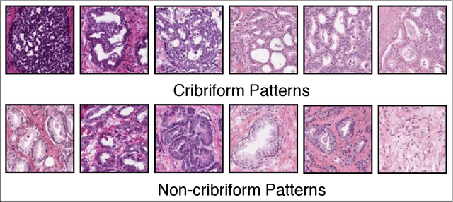
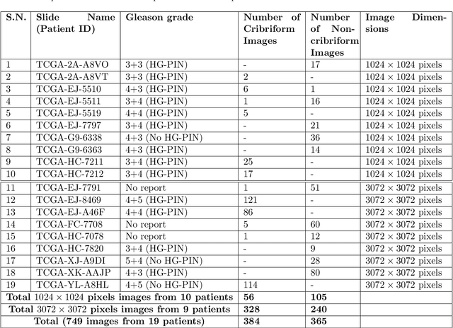
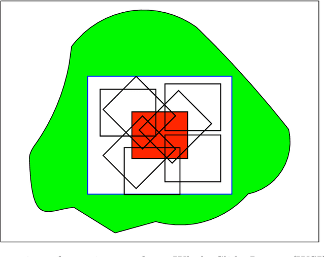
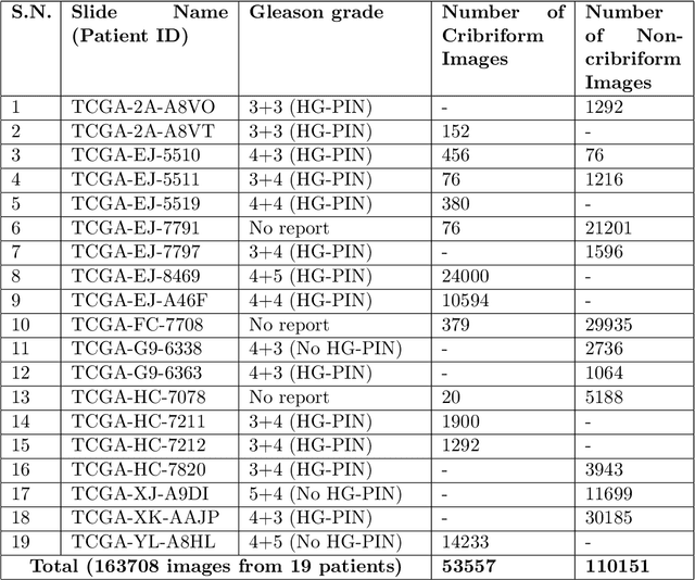
Abstract:Architecture, size, and shape of glands are most important patterns used by pathologists for assessment of cancer malignancy in prostate histopathological tissue slides. Varying structures of glands along with cumbersome manual observations may result in subjective and inconsistent assessment. Cribriform gland with irregular border is an important feature in Gleason pattern 4. We propose using deep neural networks for cribriform pattern classification in prostate histopathological images. $163708$ Hematoxylin and Eosin (H\&E) stained images were extracted from histopathologic tissue slides of $19$ patients with prostate cancer and annotated for cribriform patterns. Our automated image classification system analyses the H\&E images to classify them as either `Cribriform' or `Non-cribriform'. Our system uses various deep learning approaches and hand-crafted image pixel intensity-based features. We present our results for cribriform pattern detection across various parameters and configuration allowed by our system. The combination of fine-tuned deep learning models outperformed the state-of-art nuclei feature based methods. Our image classification system achieved the testing accuracy of $85.93~\pm~7.54$ (cross-validated) and $88.04~\pm~5.63$ ( additional unseen test set) across three folds. In this paper, we present an annotated cribriform dataset along with analysis of deep learning models and hand-crafted features for cribriform pattern detection in prostate histopathological images.
Lung Nodule Classification using Deep Local-Global Networks
Apr 23, 2019


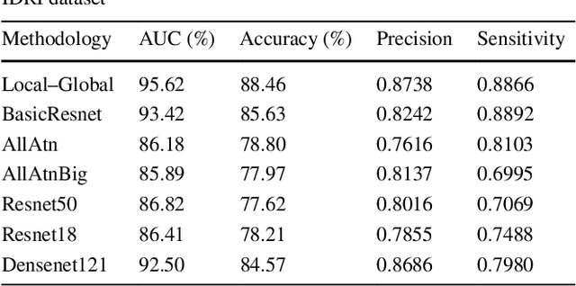
Abstract:Purpose: Lung nodules have very diverse shapes and sizes, which makes classifying them as benign/malignant a challenging problem. In this paper, we propose a novel method to predict the malignancy of nodules that have the capability to analyze the shape and size of a nodule using a global feature extractor, as well as the density and structure of the nodule using a local feature extractor. Methods: We propose to use Residual Blocks with a 3x3 kernel size for local feature extraction, and Non-Local Blocks to extract the global features. The Non-Local Block has the ability to extract global features without using a huge number of parameters. The key idea behind the Non-Local Block is to apply matrix multiplications between features on the same feature maps. Results: We trained and validated the proposed method on the LIDC-IDRI dataset which contains 1,018 computed tomography (CT) scans. We followed a rigorous procedure for experimental setup namely, 10-fold cross-validation and ignored the nodules that had been annotated by less than 3 radiologists. The proposed method achieved state-of-the-art results with AUC=95.62%, while significantly outperforming other baseline methods. Conclusions: Our proposed Deep Local-Global network has the capability to accurately extract both local and global features. Our new method outperforms state-of-the-art architecture including Densenet and Resnet with transfer learning.
Gated-Dilated Networks for Lung Nodule Classification in CT scans
Jan 01, 2019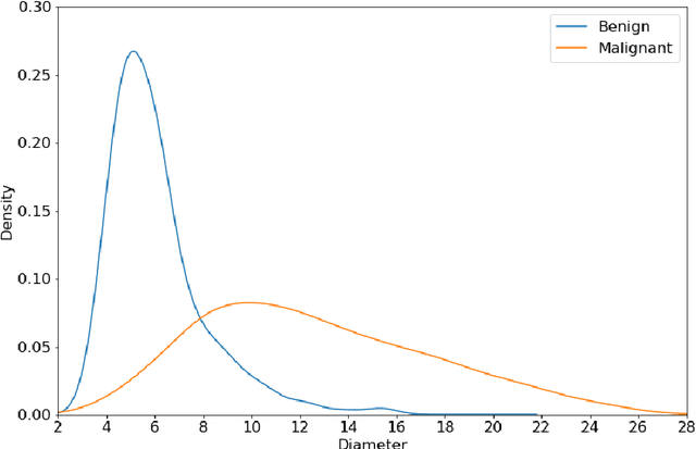

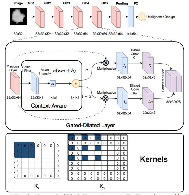

Abstract:Different types of Convolutional Neural Networks (CNNs) have been applied to detect cancerous lung nodules from computed tomography (CT) scans. However, the size of a nodule is very diverse and can range anywhere between 3 and 30 millimeters. The high variation of nodule sizes makes classifying them a difficult and challenging task. In this study, we propose a novel CNN architecture called Gated-Dilated (GD) Networks to classify nodules as malignant or benign. Unlike previous studies, the GD network uses multiple dilated convolutions instead of max-poolings to capture the scale variations. Moreover, the GD network has a Context-Aware sub-network that analyzes the input features and guides the features to a suitable dilated convolution. We evaluated the proposed network on more than 1,000 CT scans from the LIDC-LDRI dataset. Our proposed network outperforms baseline models including conventional CNNs, Resnet, and Densenet, with an AUC of >0.95. Compared to the baseline models, the GD network improves the classification accuracies of mid-range sized nodules. Furthermore, we observe a relationship between the size of the nodule and the attention signal generated by the Context-Aware sub-network, which validates our new network architecture.
 Add to Chrome
 Add to Firefox
 Add to Edge