Martin Gunn
Identifying Imaging Follow-Up in Radiology Reports: A Comparative Analysis of Traditional ML and LLM Approaches
Nov 14, 2025Abstract:Large language models (LLMs) have shown considerable promise in clinical natural language processing, yet few domain-specific datasets exist to rigorously evaluate their performance on radiology tasks. In this work, we introduce an annotated corpus of 6,393 radiology reports from 586 patients, each labeled for follow-up imaging status, to support the development and benchmarking of follow-up adherence detection systems. Using this corpus, we systematically compared traditional machine-learning classifiers, including logistic regression (LR), support vector machines (SVM), Longformer, and a fully fine-tuned Llama3-8B-Instruct, with recent generative LLMs. To evaluate generative LLMs, we tested GPT-4o and the open-source GPT-OSS-20B under two configurations: a baseline (Base) and a task-optimized (Advanced) setting that focused inputs on metadata, recommendation sentences, and their surrounding context. A refined prompt for GPT-OSS-20B further improved reasoning accuracy. Performance was assessed using precision, recall, and F1 scores with 95% confidence intervals estimated via non-parametric bootstrapping. Inter-annotator agreement was high (F1 = 0.846). GPT-4o (Advanced) achieved the best performance (F1 = 0.832), followed closely by GPT-OSS-20B (Advanced; F1 = 0.828). LR and SVM also performed strongly (F1 = 0.776 and 0.775), underscoring that while LLMs approach human-level agreement through prompt optimization, interpretable and resource-efficient models remain valuable baselines.
A Novel Corpus of Annotated Medical Imaging Reports and Information Extraction Results Using BERT-based Language Models
Mar 27, 2024
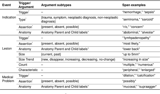
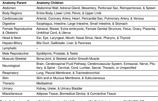
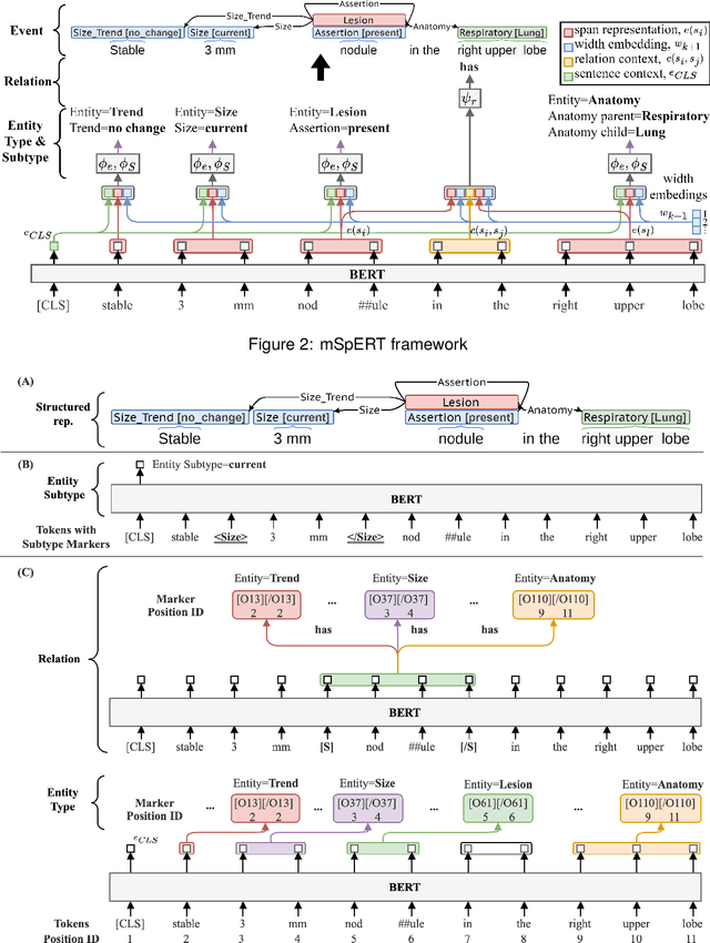
Abstract:Medical imaging is critical to the diagnosis, surveillance, and treatment of many health conditions, including oncological, neurological, cardiovascular, and musculoskeletal disorders, among others. Radiologists interpret these complex, unstructured images and articulate their assessments through narrative reports that remain largely unstructured. This unstructured narrative must be converted into a structured semantic representation to facilitate secondary applications such as retrospective analyses or clinical decision support. Here, we introduce the Corpus of Annotated Medical Imaging Reports (CAMIR), which includes 609 annotated radiology reports from three imaging modality types: Computed Tomography, Magnetic Resonance Imaging, and Positron Emission Tomography-Computed Tomography. Reports were annotated using an event-based schema that captures clinical indications, lesions, and medical problems. Each event consists of a trigger and multiple arguments, and a majority of the argument types, including anatomy, normalize the spans to pre-defined concepts to facilitate secondary use. CAMIR uniquely combines a granular event structure and concept normalization. To extract CAMIR events, we explored two BERT (Bi-directional Encoder Representation from Transformers)-based architectures, including an existing architecture (mSpERT) that jointly extracts all event information and a multi-step approach (PL-Marker++) that we augmented for the CAMIR schema.
Extracting Radiological Findings With Normalized Anatomical Information Using a Span-Based BERT Relation Extraction Model
Aug 20, 2021
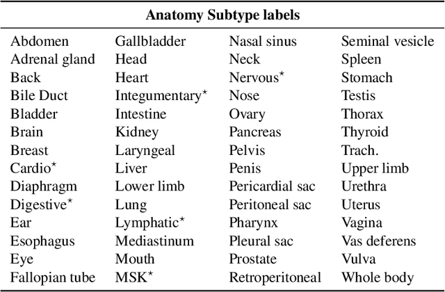

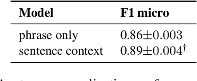
Abstract:Medical imaging is critical to the diagnosis and treatment of numerous medical problems, including many forms of cancer. Medical imaging reports distill the findings and observations of radiologists, creating an unstructured textual representation of unstructured medical images. Large-scale use of this text-encoded information requires converting the unstructured text to a structured, semantic representation. We explore the extraction and normalization of anatomical information in radiology reports that is associated with radiological findings. We investigate this extraction and normalization task using a span-based relation extraction model that jointly extracts entities and relations using BERT. This work examines the factors that influence extraction and normalization performance, including the body part/organ system, frequency of occurrence, span length, and span diversity. It discusses approaches for improving performance and creating high-quality semantic representations of radiological phenomena.
Automatic Assignment of Radiology Examination Protocols Using Pre-trained Language Models with Knowledge Distillation
Sep 01, 2020


Abstract:Selecting radiology examination protocol is a repetitive, error-prone, and time-consuming process. In this paper, we present a deep learning approach to automatically assign protocols to computer tomography examinations, by pre-training a domain-specific BERT model ($BERT_{rad}$). To handle the high data imbalance across exam protocols, we used a knowledge distillation approach that up-sampled the minority classes through data augmentation. We compared classification performance of the described approach with the statistical n-gram models using Support Vector Machine (SVM) and Random Forest (RF) classifiers, as well as the Google's $BERT_{base}$ model. SVM and RF achieved macro-averaged F1 scores of 0.45 and 0.6 while $BERT_{base}$ and $BERT_{rad}$ achieved 0.61 and 0.63. Knowledge distillation improved overall performance on the minority classes, achieving a F1 score of 0.66. Additionally, by choosing the optimal threshold, the BERT models could classify over 50% of test samples within 5% error rate and potentially alleviate half of radiologist protocoling workload.
 Add to Chrome
 Add to Firefox
 Add to Edge