 Add to Chrome
 Add to Firefox
 Add to Edge
CAHAL: Clinically Applicable resolution enHAncement for Low-resolution MRI scans
Apr 20, 2026Large-scale automated morphometric analysis of brain MRI is limited by the thick-slice, anisotropic acquisitions prevalent in routine clinical practice. Existing generative super-resolution (SR) methods produce visually compelling isotropic volumes but often introduce anatomical hallucinations, systematic volumetric overestimation, and structural distortions that compromise downstream quantitative analysis and diagnostic safety. To address this, we propose CAHAL (Clinically Applicable resolution enHAncement for Low-resolution MRI scans), a hallucination-robust, physics-informed resolution enhancement framework that operates directly in the patient's native acquisition space. CAHAL employs a deterministic bivariate Mixture of Experts (MoE) architecture routing each input through specialised residual 3D U-Net experts conditioned on both volumetric resolution and acquisition anisotropy, two independent descriptors of clinical MRI acquisition. Experts are optimised with a composite loss combining edge-penalised spatial reconstruction, Fourier-domain spectral coherence matching, and a segmentation-guided semantic consistency constraint. Training pairs are generated on-the-fly via physics-based degradation sampled from a large-scale real-world database, ensuring robust generalisation. Validated on T1-weighted and FLAIR sequences against generative baselines, CAHAL achieves state-of-the-art results, improving the best related methods in terms of accuracy and efficiency.
Ultra-high resolution multimodal MRI dense labelled holistic brain atlas
Jan 28, 2025


In this paper, we introduce holiAtlas, a holistic, multimodal and high-resolution human brain atlas. This atlas covers different levels of details of the human brain anatomy, from the organ to the substructure level, using a new dense labelled protocol generated from the fusion of multiple local protocols at different scales. This atlas has been constructed averaging images and segmentations of 75 healthy subjects from the Human Connectome Project database. Specifically, MR images of T1, T2 and WMn (White Matter nulled) contrasts at 0.125 $mm^{3}$ resolution that were nonlinearly registered and averaged using symmetric group-wise normalisation to construct the atlas. At the finest level, the holiAtlas protocol has 350 different labels derived from 10 different delineation protocols. These labels were grouped at different scales to provide a holistic view of the brain at different levels in a coherent and consistent manner. This multiscale and multimodal atlas can be used for the development of new ultra-high resolution segmentation methods that can potentially leverage the early detection of neurological disorders.
DeepCERES: A Deep learning method for cerebellar lobule segmentation using ultra-high resolution multimodal MRI
Jan 23, 2024


This paper introduces a novel multimodal and high-resolution human brain cerebellum lobule segmentation method. Unlike current tools that operate at standard resolution ($1 \text{ mm}^{3}$) or using mono-modal data, the proposed method improves cerebellum lobule segmentation through the use of a multimodal and ultra-high resolution ($0.125 \text{ mm}^{3}$) training dataset. To develop the method, first, a database of semi-automatically labelled cerebellum lobules was created to train the proposed method with ultra-high resolution T1 and T2 MR images. Then, an ensemble of deep networks has been designed and developed, allowing the proposed method to excel in the complex cerebellum lobule segmentation task, improving precision while being memory efficient. Notably, our approach deviates from the traditional U-Net model by exploring alternative architectures. We have also integrated deep learning with classical machine learning methods incorporating a priori knowledge from multi-atlas segmentation, which improved precision and robustness. Finally, a new online pipeline, named DeepCERES, has been developed to make available the proposed method to the scientific community requiring as input only a single T1 MR image at standard resolution.
Machine learning approaches for COVID-19 detection from chest X-ray imaging: A Systematic Review
Jun 11, 2022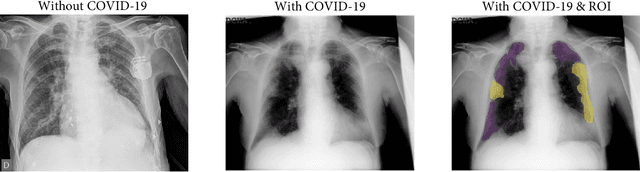
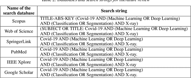

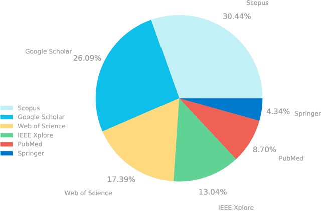
There is a necessity to develop affordable, and reliable diagnostic tools, which allow containing the COVID-19 spreading. Machine Learning (ML) algorithms have been proposed to design support decision-making systems to assess chest X-ray images, which have proven to be useful to detect and evaluate disease progression. Many research articles are published around this subject, which makes it difficult to identify the best approaches for future work. This paper presents a systematic review of ML applied to COVID-19 detection using chest X-ray images, aiming to offer a baseline for researchers in terms of methods, architectures, databases, and current limitations.